
Diffusion Tensor Imaging Collection
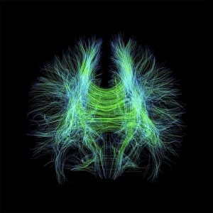
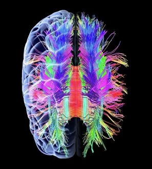
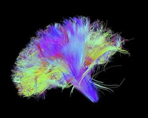
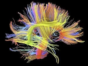
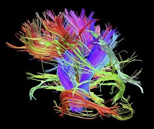
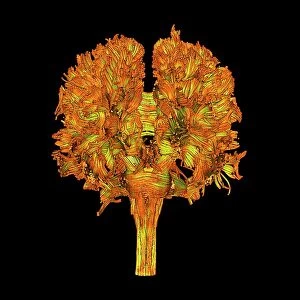
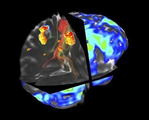
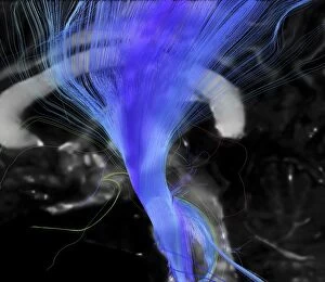
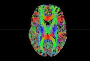
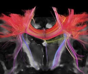
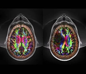
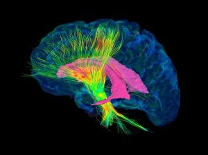
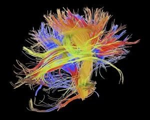
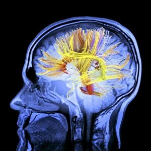
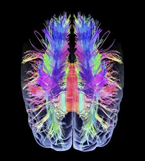
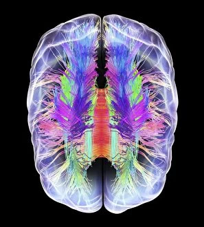
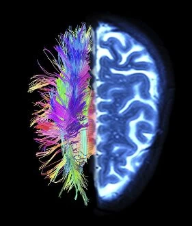
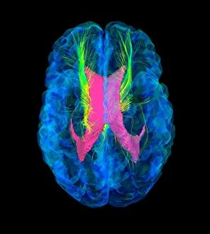
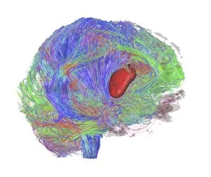
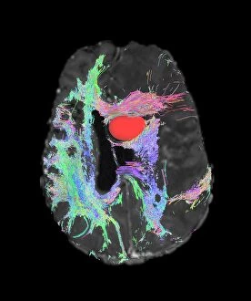
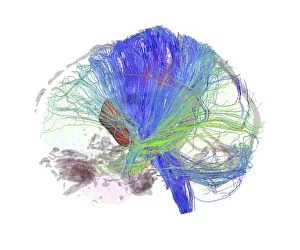
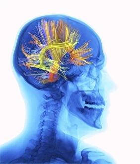
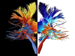
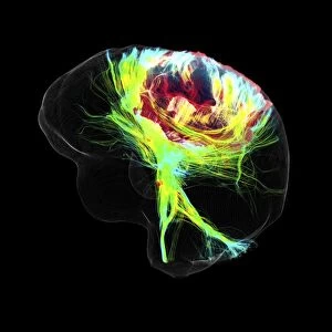
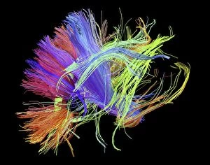
Diffusion tensor imaging (DTI) is a revolutionary technique that allows us to visualize and understand the intricate network of brain fibres


For sale as Licensed Images
Choose your image, Select your licence and Download the media
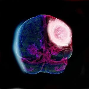
Diffusion tensor imaging (DTI) is a revolutionary technique that allows us to visualize and understand the intricate network of brain fibres. Using an MRI scan, such as C017 / 7099 or C017 / 7035, DTI provides valuable insights into the white matter fibres that connect different regions of the human brain. The artwork C015 / 1930 beautifully depicts these white matter fibres and their complex arrangement within our brains, and is truly fascinating how these delicate structures enable efficient communication between various brain regions, facilitating our cognitive processes. C014 / 5666, C014 / 5668, and C014 / 5667 showcase the mesmerizing intricacy of white matter fibres in the human brain. These images highlight the importance of understanding this vast network for unraveling mysteries related to cognition, behavior, and neurological disorders. By utilizing DTI scans like those depicted in "White Matter Fibers - DTI Scan, " researchers can delve deeper into studying conditions such as brain tumors (as seen in fMRI and tractography image C017 / 7102). This advanced technology aids in mapping out critical areas affected by tumors while preserving essential functions during surgical planning. Furthermore, DTI MRI scan C017/7046 illustrates the corticospinal tract's significance—a crucial pathway responsible for motor function—allowing us to comprehend its role better in movement-related disorders or injuries. Tract density imaging (TDI) offers another perspective on brain fibers' distribution, as shown in image C017/7039. By examining TDI scans alongside traditional DTIs, we gain a comprehensive understanding of fiber connectivity throughout the brain. DTI modeling plays a pivotal role when investigating brain tumors (as demonstrated by image C017/7060). This technique helps create accurate models that aid surgeons in navigating around vital structures during tumor resection surgeries while minimizing damage to healthy tissue.