Digestive System Collection (#31)

The digestive system is a complex network of organs and processes that allows our bodies to break down food and absorb nutrients

Background imageDigestive System Collection: Abdominal organs

Abdominal organs, historical anatomical artwork. This ventral (front) view of a dissected abdomen shows several of the organs involved in digestion. The stomach has been removed

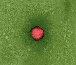
Background imageDigestive System Collection: Adenovirus, TEM

Adenovirus, TEM

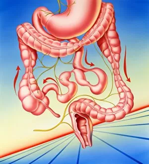
Background imageDigestive System Collection: Irritable bowel syndrome

Irritable bowel syndrome. Artwork of a digestive system suffering from irritable bowel syndrome (IBS). IBS is also known as spastic colon or irritable colon syndrome

Background imageDigestive System Collection: Gases in the abdomen, X-ray

Gases in the abdomen, X-ray
Bloated abdomen in tympanites, coloured X-ray. Tympanites is swelling of the abdomen due to the accumulation of gases in the intestines or the peritoneal cavity. Gas pockets are seen as dark spaces

Background imageDigestive System Collection: Hiatus hernia, X-ray

Hiatus hernia, X-ray
Hiatus hernia, coloured X-ray. Side view of a patient suffering from a hiatus hernia, the protrusion of the stomach through the diaphragm into the chest

Background imageDigestive System Collection: Artwork of digestive system and harmful bacteria

Artwork of digestive system and harmful bacteria
Gastroenteritis. Artwork of bacteria shown below the human digestive system in which they cause gastroenteritis, the inflammation of the stomach and intestines

Background imageDigestive System Collection: Ulcerative colitis, SEM

Ulcerative colitis, SEM
Ulcerative colitis. Coloured scanning electron micrograph (SEM) of freeze-fractured bowel tissue from a patient suffering from ulcerative colitis

Background imageDigestive System Collection: Normal intestines, 3D CT scan

Normal intestines, 3D CT scan
Normal intestines. Coloured 3D computed tomography (CT) scan of the healthy intestines of 38 year old patient

Background imageDigestive System Collection: Earthworm gut, light micrograph

Earthworm gut, light micrograph
Earthworm. Light micrograph of a section through the gut of an earthworm (Lumbricus terrestris). Magnification: x15 when printed at 10 centimetres wide

Background imageDigestive System Collection: Abdominal anatomy, artwork

Abdominal anatomy, artwork
Abdominal anatomy, computer artwork

Background imageDigestive System Collection: Megacolon, artwork

Megacolon, artwork
Megacolon, computer artwork. This is an abnormal dilation of the colon that may be congenital or acquired

Background imageDigestive System Collection: Healthy stomach, artwork

Healthy stomach, artwork

Background imageDigestive System Collection: Healthy liver, artwork

Healthy liver, artwork

Background imageDigestive System Collection: Healthy large intestines, artwork

Healthy large intestines, artwork

Background imageDigestive System Collection: Healthy intestines, artwork

Healthy intestines, artwork

Background imageDigestive System Collection: Healthy large intestine, artwork

Healthy large intestine, artwork

Background imageDigestive System Collection: Hookworm head, artwork

Hookworm head, artwork

Background imageDigestive System Collection: Stomach lining with gastric pits, SEM

Stomach lining with gastric pits, SEM
Stomach lining. Coloured scanning electron micrograph (SEM) of the inner lining of the stomach (gastric mucosa). The large folds are known as rugae

Background imageDigestive System Collection: Intestinal villi, artwork

Intestinal villi, artwork

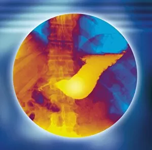
Background imageDigestive System Collection: Normal stomach, X-ray

Normal stomach, X-ray
Normal stomach (centre), coloured frontal X-ray. The stomach has been highlighted by an ingested barium meal. The spine is seen running from top to bottom

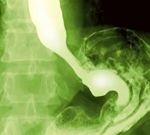
Background imageDigestive System Collection: Normal oesophagus and stomach, X-ray

Normal oesophagus and stomach, X-ray
Normal oesophagus and stomach. Coloured frontal X-ray of a healthy oesophagus (top left) and stomach (lower right), which have been highlighted by ingested barium meal

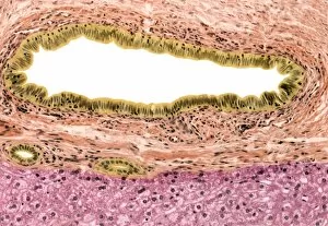
Background imageDigestive System Collection: Bile duct, light micrograph

Bile duct, light micrograph. Microvilli are brown, columnar epithelium is orange. Connective tissue is pink. Two small bile ducts are also seen towards bottom left

Background imageDigestive System Collection: Colon lining, light micrograph

Colon lining, light micrograph
Colon lining. Light micrograph of a section the lining of the colon, highlighting the mucous producing goblet cells (purple). Magnification: x200 when printed at 10 centimetres wide

Background imageDigestive System Collection: Stomach lining, light micrograph

Stomach lining, light micrograph. The stomach lumen is lined by mucous cells. At bottom centre is an accumulation of lymphocytes forming a lymphatic nodule in the sub mucosa


For sale as Licensed Images

Choose your image, Select your licence and Download the media

The digestive system is a complex network of organs and processes that allows our bodies to break down food and absorb nutrients. From the gastrointestinal nematodes that inhabit our intestines to the medical illustration of an appendix with appendicitis, every component plays a vital role in this intricate system. Bee anatomy, depicted through artwork, showcases how these tiny creatures have their own unique digestive systems. Meanwhile, a scanning electron microscope (SEM) image reveals the Salmonella bacteria responsible for causing foodborne illnesses. Another SEM image focuses on the small intestine's surface, highlighting its crucial function in nutrient absorption. Similarly, we see the gallbladder's surface under SEM examination, emphasizing its role in storing bile produced by the liver. C. Elegans worms are also featured here as they provide valuable insights into understanding human digestion at a microscopic level. A light micrograph captures their intricate structure and behavior within this fascinating ecosystem. Moving beyond microscopic organisms, we encounter a muskrat sitting on the shore of a pond indulging in grass consumption—an example of herbivorous digestion within nature's cycle. An X-ray image provides us with an inside look at an inflamed appendix—a condition known as appendicitis—underscoring both its importance and potential health risks if left untreated. Lastly, artwork showcasing pancreas anatomy reminds us of this organ's significant contribution to digestion through hormone production such as insulin regulation.