
Digestive Collection (#3)
From McVitie's Digestive in 1936 to Enos Salts in 1897, digestive aids have been a part of our lives for centuries


For sale as Licensed Images
Choose your image, Select your licence and Download the media
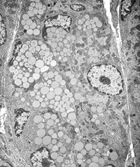
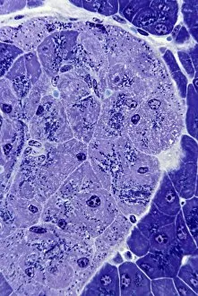
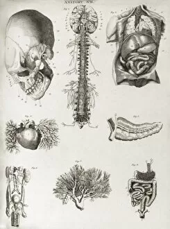
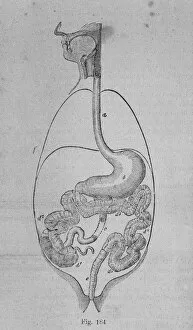
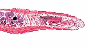
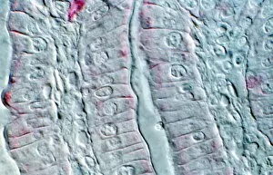
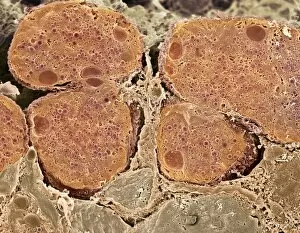
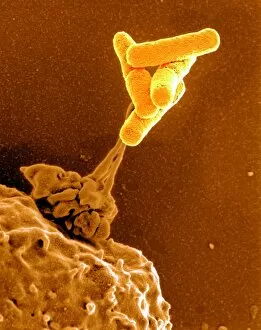
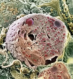
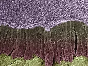
From McVitie's Digestive in 1936 to Enos Salts in 1897, digestive aids have been a part of our lives for centuries. The iconic McVitie's Digestive biscuit, introduced in 1934 by McVitie & Price, has become a beloved treat enjoyed with tea or on its own. Meanwhile, Enos Salts made their debut in 1897 and quickly gained popularity as an effective remedy for indigestion. Enos Cockerel became the face of Enos Fruit Salts through captivating advertisements that promised relief from digestive discomfort. These ads featured vibrant illustrations and catchy slogans that caught the attention of consumers. Understanding our digestive organs is crucial to maintaining good health. Just like the intricate fish anatomy depicted in historical models, our own intestines play a vital role in breaking down food and absorbing nutrients. However, they can also be susceptible to intestinal protozoan parasites as shown under the microscope using TEM technology. Even animals benefit from digestive aids. Artwork depicting dog anatomy showcases how similar their systems are to ours when it comes to digestion. It reminds us that taking care of our furry friends' stomachs is just as important as taking care of our own. In moments of reflection, we find even spiritual figures like Buddha acknowledging the importance of digestion. As history tells us, Buddha passed away after consuming contaminated pork – a stark reminder that proper digestion is essential for overall well-being. Advertisements throughout time have highlighted various products aimed at improving digestion. One such example is Pruna - a color litho ad featuring mouthwatering fruits encourages consumers to indulge while ensuring optimal digestion. Whether it's enjoying a delicious McVitie's Digestive biscuit or finding relief with Enos Salts, these hints remind us that caring for our digestive system has always been an integral part of human history.