
Electron Micrograph Collection (#5)
"Unlocking the Hidden World: Exploring Electron Micrographs" Delve into the microscopic realm and witness the intricate beauty of life through electron micrographs


For sale as Licensed Images
Choose your image, Select your licence and Download the media
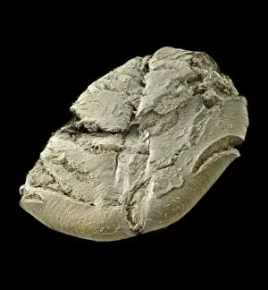
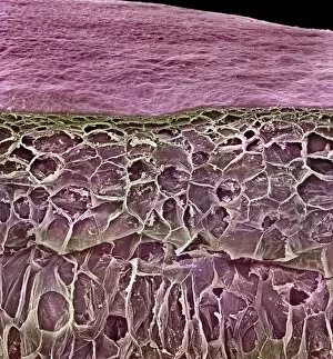
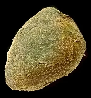
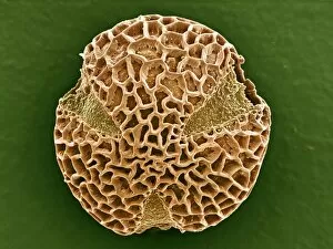
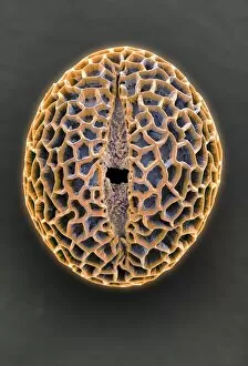
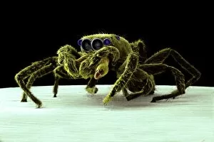
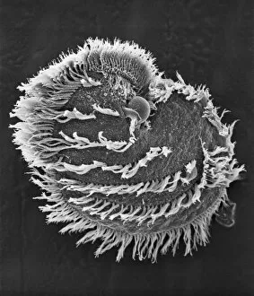
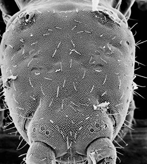
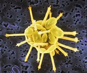
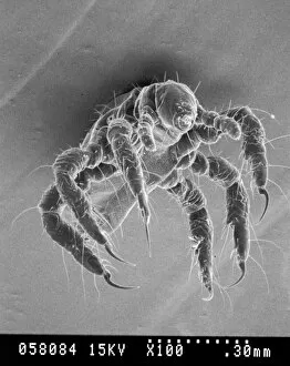
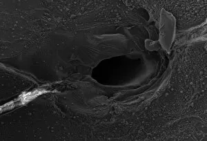
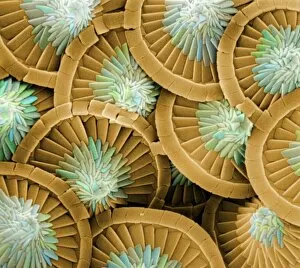
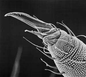
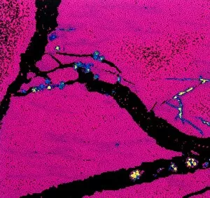
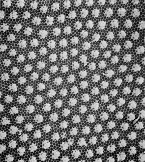
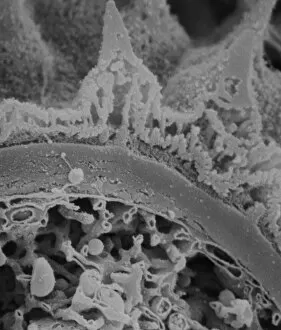
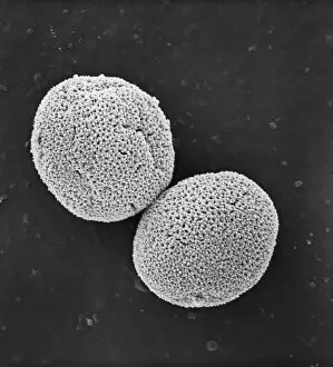
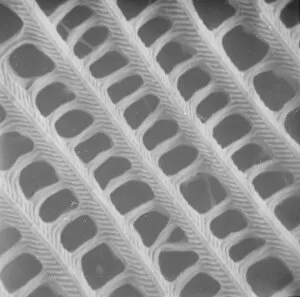
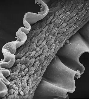
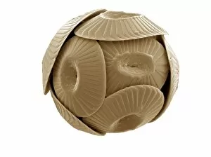
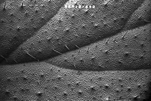
"Unlocking the Hidden World: Exploring Electron Micrographs" Delve into the microscopic realm and witness the intricate beauty of life through electron micrographs. Discosphaera tubifera, coccolithophore: Marvel at the stunning calcium carbonate plates adorning this marine phytoplankton, resembling a delicate work of art. Crysotile asbestos: Peer into the dangerous fibers that make up this mineral, revealing its hazardous nature when inhaled. Liver: Journey inside this vital organ and observe its complex network of cells, unveiling its role in detoxification and metabolism. Cimex lectularius, bed bug: Get up close with these notorious pests as their exoskeletons reveal their resilience to survive even against our best efforts. Coloured TEM of Yersinia pestis bacteria: Witness the haunting beauty of these deadly bacteria responsible for causing plague outbreaks throughout history. Taraxacum officinale, dandelion (fruiting head): Explore the intricate structure of a dandelion's fruiting head under high magnification, showcasing nature's ingenious method for seed dispersal. Simulium damnosum, Simulian blackfly: Encounter these tiny insects known for transmitting river blindness as you uncover their detailed anatomy and feeding mechanisms. Norovirus particles, TEM: Enter the world of viruses as you observe norovirus particles - a common cause of gastrointestinal illness - providing insights into their structure and potential vulnerabilities for future treatments. 9 & 10 E. coli bacterium/bacteria : Dive deep into both individual E. coli cells or colonies to understand their role in digestion while also highlighting concerns surrounding foodborne illnesses caused by certain strains. Snail teeth : Discover how snails possess an unexpected weapon – razor-sharp teeth – enabling them to feed on tough plant material with ease; an evolutionary marvel. Chloroplast in cell of pea plant.