
Electron Microscope Collection (#3)
The electron microscope, a marvel of scientific innovation, has revolutionized our understanding of the microscopic world


For sale as Licensed Images
Choose your image, Select your licence and Download the media
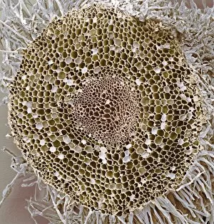
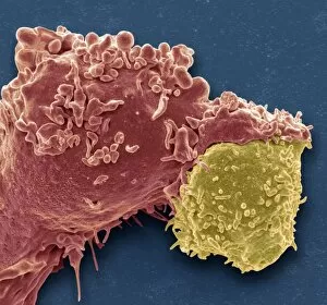
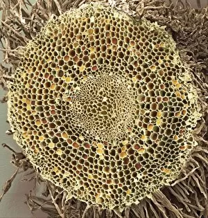
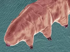
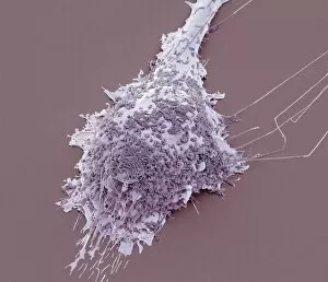
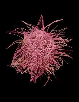
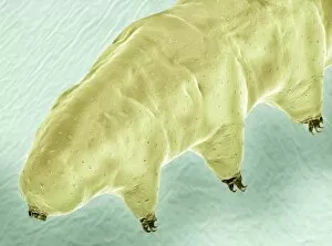
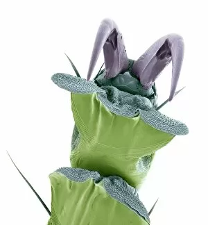
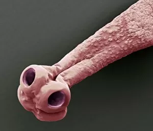
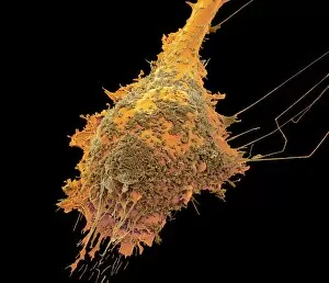
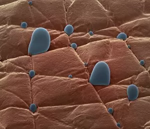
The electron microscope, a marvel of scientific innovation, has revolutionized our understanding of the microscopic world. With its incredible magnification capabilities, it allows us to delve into the intricate details of various specimens and uncover hidden wonders. One such specimen is the osteocyte bone cell, captured in stunning detail by SEM C016 / 9025. This image reveals the complex structure and delicate nature of these cells that play a crucial role in maintaining healthy bones. In another captivating image (SEM C016 / 9084), we are introduced to the water bear, an extraordinary creature known for its ability to survive extreme conditions. Through the electron microscope's lens, we witness its unique anatomy and gain insight into how it adapts to different environments. Moving on from living organisms, we explore Emiliania huxleyi coccolithophores – tiny marine algae with intricately patterned shells. The electron microscope unveils their exquisite beauty and provides valuable information about their ecological significance, and is worth noting that this remarkable instrument owes its existence to companies like Zeiss from Jena who have dedicated themselves to pushing technological boundaries in microscopy. Their contributions have paved the way for groundbreaking discoveries across various scientific disciplines. Looking back through history, we encounter an engraving depicting Robert Koch in his laboratory during the 19th century. This iconic image reminds us of how far microscopy has come since then and highlights Koch's influential work in microbiology. Additionally, we are transported back over a century ago with photographs showcasing studies on snowflakes' form and structure around 1902-1903. These mesmerizing images capture nature's artistry at its finest while demonstrating how electron microscopes enable us to unravel even seemingly simple phenomena like snowflakes' intricate designs. The electron microscope continues to amaze scientists and enthusiasts alike with its ability to reveal hidden worlds beyond our naked eye's reach.