Epithelial Collection (#6)

Epithelial cells, the unsung heroes of our body's lining

Background imageEpithelial Collection: Intestinal villus cell loss, SEM

Intestinal villus cell loss, SEM
Intestinal villus cell loss, coloured scanning electron micrograph (SEM). Close-up of the tip of a villus in the small intestine that has shed a cell from its tip as part of the normal cell

Background imageEpithelial Collection: Intestinal villi cell loss, SEM

Intestinal villi cell loss, SEM
Intestinal villi cell loss, coloured scanning electron micrograph (SEM). Close-up of the tips of villi in the small intestine

Background imageEpithelial Collection: Meninges, artwork

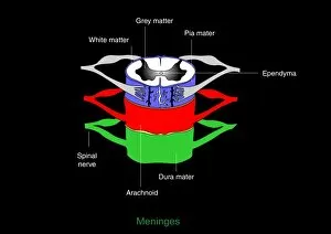
Meninges, artwork
Meninges. Computer artwork showing the meninges surrounding the spinal cord. The meninges are the three membranes that envelop the brain and spinal cord

Background imageEpithelial Collection: Blood vessel, artwork

Blood vessel, artwork
Blood vessel. Computer artwork of the inside of a blood vessel, showing red blood cells (circular) and the epithelial cells linging the vessels wall

Background imageEpithelial Collection: Fallopian tube, SEM

Fallopian tube, SEM
Fallopian tube. Coloured scanning electron micrograph (SEM) of the lining of a fallopian tube (oviduct). The fallopian tubes carry the egg from the ovary to the uterus (womb)

Background imageEpithelial Collection: Nasal mucosa, artwork

Nasal mucosa, artwork
Nasal mucosa, cross-section. Artwork of a sequence (left to right) showing mucus production in response to infection and inflammation

Background imageEpithelial Collection: Lung cells, fluorescent micrograph

Lung cells, fluorescent micrograph
Lung cells. Immunofluorescence light micrograph of pulmonary endothelial cells. Endothelial cells are specialized epithelial cells that line the inner surface of blood vessels

Background imageEpithelial Collection: Olfactory epithelium, artwork

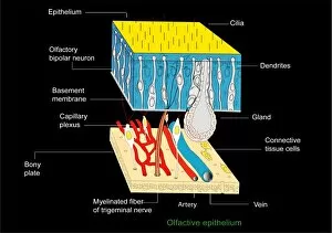
Olfactory epithelium, artwork
Olfactory epithelium. Computer artwork showing the structure of the specialised layer of tissue that lines the inside of the nasal cavity and is involved in smell

Background imageEpithelial Collection: Small intestine lining, light micrograph

Small intestine lining, light micrograph
Small intestine lining. Light micrograph of a section through the finger-like projections (villi) of the duodenum, the uppermost part of the small intestine

Background imageEpithelial Collection: Gall bladder surface, light micrograph

Gall bladder surface, light micrograph
Gall bladder surface. Coloured light micrograph of a section through a gall bladder, showing the surface tissue layers. The gall bladders surface is made up of tiny finger-like projections called

Background imageEpithelial Collection: Epithelial gonorrhoea infection, SEM

Epithelial gonorrhoea infection, SEM
Epithelial gonorrhoea infection. Coloured scanning electron micrograph (SEM) of Neisseria gonorrhoeae bacteria (clusters of small round cells) and an epithelial cell (large structure, centre)

Background imageEpithelial Collection: Bacteria in the nose, SEM

Bacteria in the nose, SEM
Bacteria in the nose. Coloured scanning electron micrograph (SEM) of bacteria (red) on the surface of the nasal cavity. One of the squamous epithelium cells (lower centre to lower right)

Background imageEpithelial Collection: Gonorrhoea bacteria, TEM

Gonorrhoea bacteria, TEM
Gonorrhoea bacteria. Coloured transmission electron micrograph (TEM) of a Neisseria gonorrhoeae bacteria diplococci (pair of cells, light orange) infecting a human epithelial cell (dark orange)

Background imageEpithelial Collection: Skin tissue, SEM

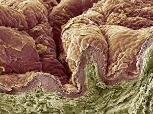
Skin tissue, SEM
Skin tissue. Coloured scanning electron micrograph (SEM) of a freeze-fracture through human skin tissue. The fracture plane (lower frame) has revealed the pseudo-stratified epithelium (below surface)

Background imageEpithelial Collection: Tooth enamel formation, SEM

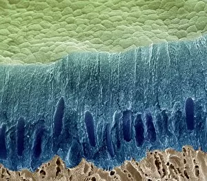
Tooth enamel formation, SEM
Tooth enamel formation. Coloured scanning electron micrograph (SEM) of a freeze-fractured section through a tooth, showing the enamel-forming cell layer (blue)

Background imageEpithelial Collection: Pancreas acinus, SEM

Pancreas acinus, SEM
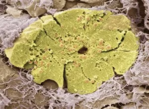
Pancreas acinus. Coloured scanning electron micrograph (SEM) of a freeze-fracture through an acinus (yellow) in the pancreas. An acinus is a collection of glandular epithelial cells

Background imageEpithelial Collection: Hyaluronic acid, molecular model

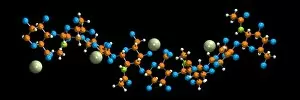
Hyaluronic acid, molecular model. Hyaluronic acid (or hyaluronon) is a glycosaminoglycan, a type of biological polymer made up of repeating units of a disaccharide (two sugar molecules)

Background imageEpithelial Collection: Dog tongue tissue, light micrograph

Dog tongue tissue, light micrograph
Dog tongue tissue. Light micrograph of a transverse section through the tongue of a dog, showing the tongues surface (across top)

Background imageEpithelial Collection: Cat tongue tissue, light micrograph

Cat tongue tissue, light micrograph
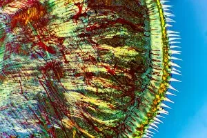
Cat tongue tissue. Polarised light micrograph of a transverse section through a cats tongue. The outer stratified epithelium (right) has spike-like (white) keratinised papillae

Background imageEpithelial Collection: Tongue tissue, light micrograph

Tongue tissue, light micrograph
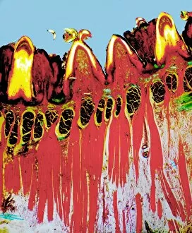
Tongue tissue. Polarised light micrograph of a transverse section through a tongue, showing the surface (across top). Here, there are four rounded fungiform papillae


For sale as Licensed Images

Choose your image, Select your licence and Download the media

Epithelial cells, the unsung heroes of our body's lining. From the intestinal tract to the trachea, these microscopic warriors protect and support us in ways we often overlook. Take a closer look at the intestinal lining under a scanning electron microscope (SEM). The intricate network of microvilli reveals itself, enhancing nutrient absorption and ensuring our digestive system functions seamlessly. Moving on to the trachea lining, SEM unveils its unique structure. These epithelial cells form a protective barrier against harmful particles while allowing for efficient gas exchange within our respiratory system. Zooming into zebra fish skin through SEM, we discover another fascinating aspect cells. Their arrangement provides defense against external threats while maintaining essential functions like osmoregulation. Intriguingly captured by SEM are bacteria residing in our nasal passages. Though seemingly insignificant, these tiny organisms play crucial roles in maintaining healthy immune responses and preventing infections. However, not all stories about epithelial cells are positive. A colour lithograph depicts the devastating impact of cancerous growth on half of someone's face. This serves as a reminder that even resilient epithelial layers can succumb to destructive forces if left unchecked. A cross-section diagram further illustrates how cancer affects various components such as calcium deposits, blood vessels, ulcerated areas, nerve fibers - all intertwined with an afflicted epithelial layer. It emphasizes the importance of early detection and treatment options available for those battling this formidable disease. Returning to less dire scenarios captured by SEM is the nasal lining – yet another example showcasing how delicate but vital these cellular linings truly are. The intestinal villi also make their appearance under SEM – finger-like projections that increase surface area for optimal nutrient absorption within our gut ecosystem. Shifting gears towards throat cancer diagnosis using X-rays highlights how medical imaging techniques aid in identifying abnormalities affecting this critical part of our anatomy. Early detection can significantly improve prognosis and treatment outcomes for patients facing such challenges.