
Epithelium Collection
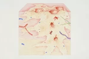
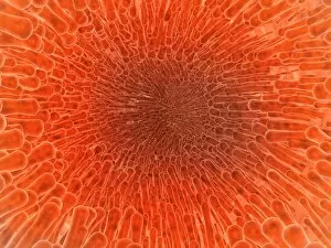
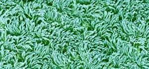
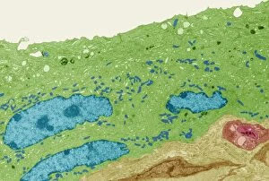
Epithelium: Unveiling the Intricacies of Our Body's Protective Shield Uterus lining during menstruation


For sale as Licensed Images
Choose your image, Select your licence and Download the media
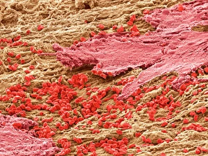
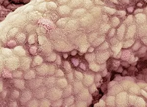
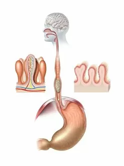
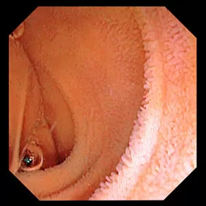
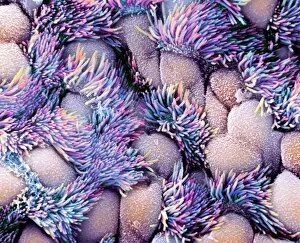
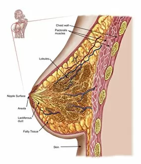
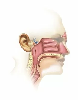
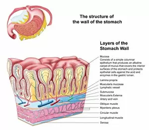
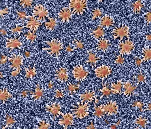
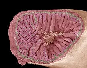
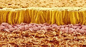
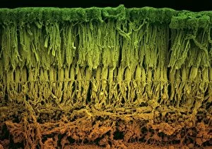
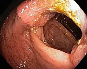
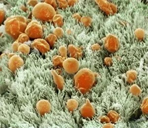
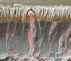
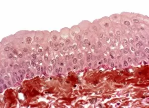
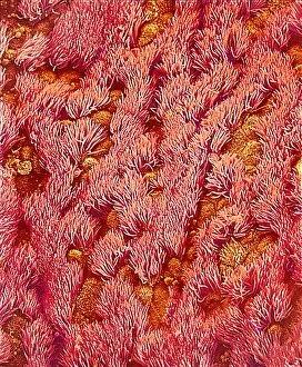
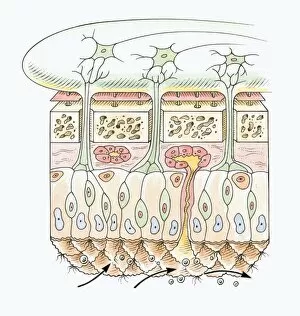
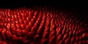
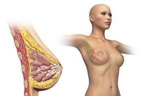
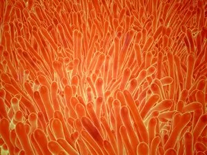
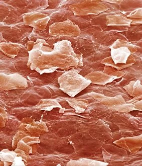
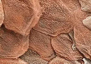
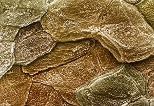
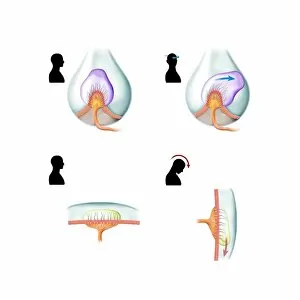
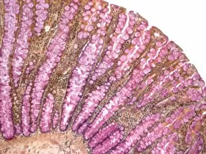
Epithelium: Unveiling the Intricacies of Our Body's Protective Shield Uterus lining during menstruation, SEM: Witness the remarkable transformation of the uterus lining during menstruation under the scanning electron microscope (SEM). Marvel at its ability to regenerate and prepare for new beginnings. Gallbladder surface, SEM: Delve into the intricate landscape of the gallbladder surface through a captivating scanning electron microscope (SEM) image. Explore how this epithelial layer aids in digestion and bile storage. Human digestive system, artwork: Embark on a visual journey through an artistic representation of our complex human digestive system. Discover how epithelial cells form protective barriers that facilitate nutrient absorption and waste elimination. Intestinal lining: Uncover the secrets held within our intestinal lining as it safeguards against harmful substances while allowing vital nutrients to pass through. Appreciate its role in maintaining overall gut health. Trachea lining, SEM: Peer into the microscopic world of tracheal epithelium using a scanning electron microscope (SEM). Observe how these specialized cells protect our airways from foreign particles and ensure smooth breathing. F. colour SEM of uterine lining, endometrium: Experience an explosion of vibrant colors as we explore the uterine lining's endometrium under a field emission color scanning electron microscope (F. colour SEM). Witness nature's breathtaking artistry up close. Intestinal microvilli, TEM: Dive deep into our intestinal microvilli with transmission electron microscopy (TEM), revealing their finger-like projections responsible for maximizing nutrient absorption efficiency – truly nature's ingenious design. Zebra fish skin, SEM: Immerse yourself in mesmerizing patterns etched onto zebra fish skin when observed under a scanning electron microscope (SEM). Admire how epithelial cells contribute to both protection and beauty in aquatic life. Bacteria in the nose, SEM.