
Escherichia Coli Collection (page 2)
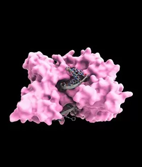
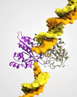
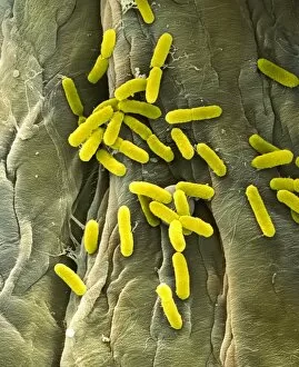
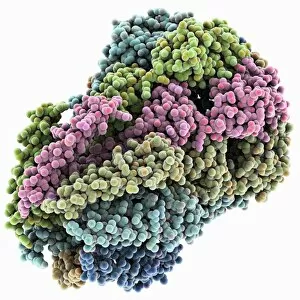
Escherichia coli, commonly known as E. Coli bacteria, is a fascinating microorganism that has captured the attention of scientists and researchers worldwide


For sale as Licensed Images
Choose your image, Select your licence and Download the media
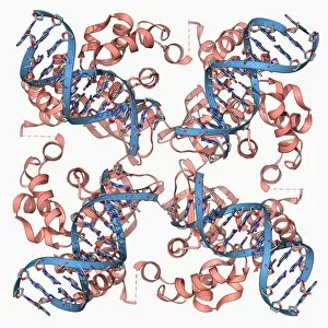
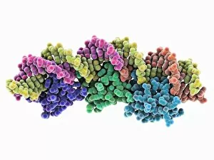
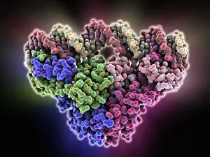
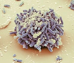
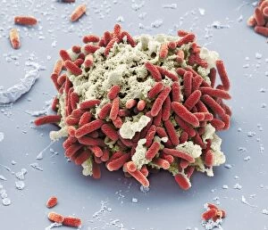
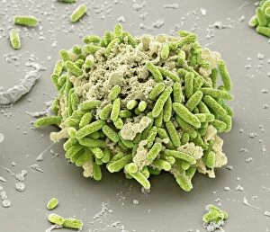
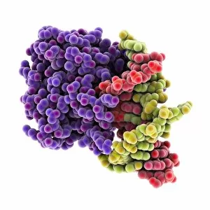
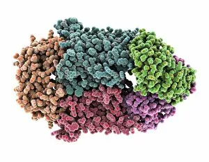
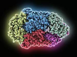
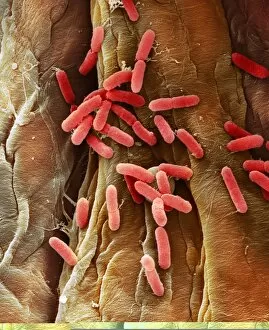
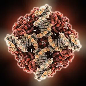
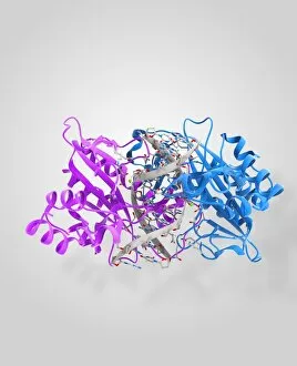
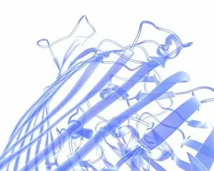
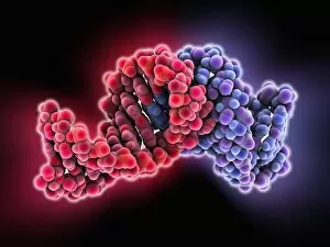
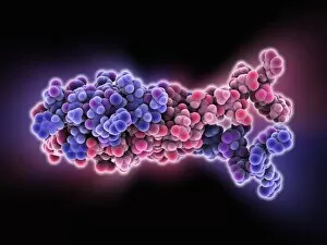
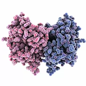
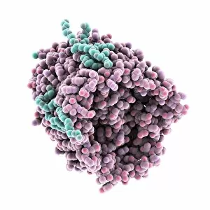
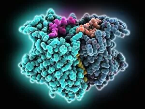
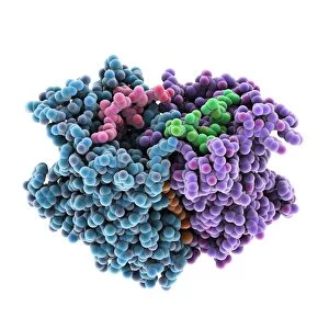
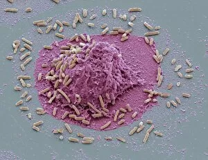
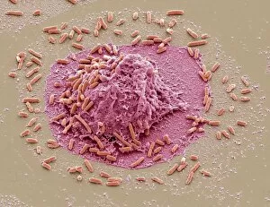
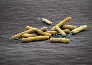
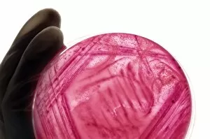
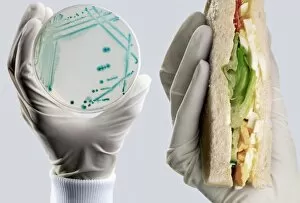
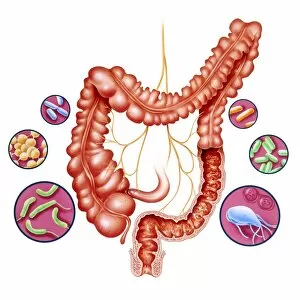
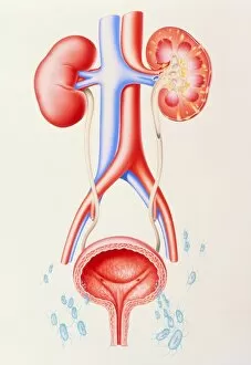
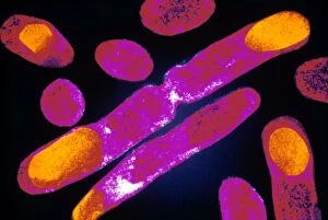
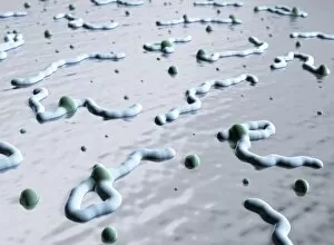
Escherichia coli, commonly known as E. Coli bacteria, is a fascinating microorganism that has captured the attention of scientists and researchers worldwide. With the help of advanced imaging techniques such as scanning electron microscopy (SEM) and transmission electron microscopy (TEM), we have been able to gain valuable insights into its structure and behavior. In SEM images, E. Coli bacteria appear as rod-shaped cells with distinct features on their surface. These tiny organisms are part of the normal flora in our intestines but can also cause various infections when they enter other parts of our body. One such infection is bladder infection, where E. Coli bacterium can be found adhering to the lining of the urinary tract. Under TEM, we get a closer look at the internal structure of this bacterium. The intricate details reveal its cell wall, cytoplasmic contents, and even its division process – a remarkable sight indeed. Additionally, specific strains like E. coli 0157: H7 have gained notoriety due to their ability to produce toxins called Shiga toxins which can lead to severe illness. To aid in visualizing these microscopic wonders more vividly, false-color TEM images provide an artistic representation while still maintaining scientific accuracy. This allows us to appreciate both the beauty and complexity hidden within this tiny world. Studying Escherichia coli is crucial for understanding bacterial pathogenesis and developing effective treatments against related infections. By unraveling its secrets through advanced imaging techniques like SEM and TEM, scientists continue striving towards improving public health by combating this versatile yet potentially harmful microbe.