
Exoskeleton Collection (#6)
"Unleashing the Power of Exoskeletons: From Ancient Defenses to Futuristic Innovations" Ankylosaurus dinosaurs defend themselves against a T-Rex


For sale as Licensed Images
Choose your image, Select your licence and Download the media
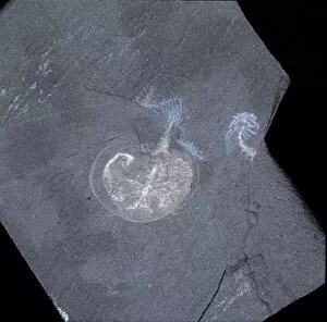
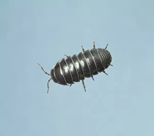
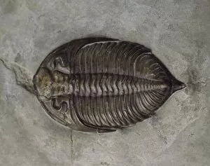
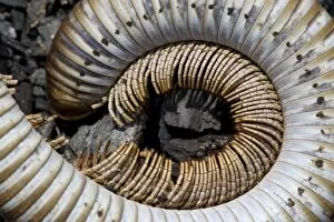
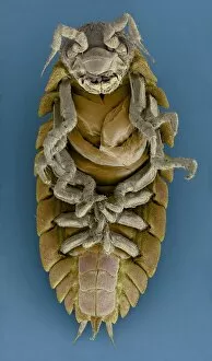
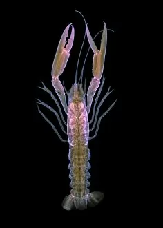
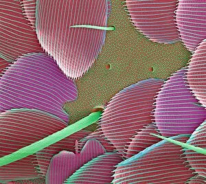
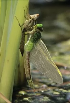
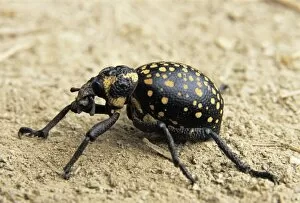
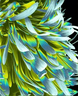
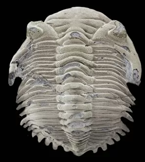
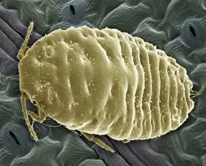
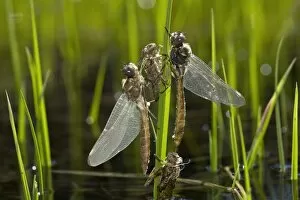
"Unleashing the Power of Exoskeletons: From Ancient Defenses to Futuristic Innovations" Ankylosaurus dinosaurs defend themselves against a T-Rex: Witness the incredible strength and resilience of these prehistoric creatures as they utilize their exoskeleton armor to ward off predators. 3D rendering of an Ankylosaurus dinosaur skeleton: Explore the intricate details of this ancient creature's exoskeletal structure, marveling at its ability to provide both protection and mobility. Insects, c1910. Creator: Unknown: Discover how insects have perfected the art of exoskeletons for millions of years, enabling them to survive in diverse environments with unmatched adaptability. Calymene blumenbachii brongniart, trilobite: Step back in time and observe the fossilized remains of a trilobite, showcasing its beautifully preserved exoskeleton that once allowed it to thrive in Earth's primordial seas. AI, AR, arms outstretched - Embracing a new era where technology meets biology; witness cutting-edge advancements in artificial intelligence and augmented reality merging seamlessly with human-like exoskeletal enhancements for enhanced balance and coordination. Black Death rat flea artwork: Uncover a dark chapter in history as we delve into the role played by fleas equipped with their own tiny yet formidable exoskeletons during one of humanity's deadliest pandemics. Spiny spider SEM image: Zoom into nature's engineering masterpiece as you explore an up-close view of a spiny spider's intricately designed exoskeleton through scanning electron microscopy (SEM). Skeleton of a seahorse (Hippocampus sp. ) showing remarkable bone structure: Dive beneath the waves and admire how even delicate creatures like seahorses possess unique skeletal structures within their protective external shells – an exquisite example of nature's ingenuity. Android, blue background, body armor.