
Extremophile Collection
"Exploring the Unseen World: Extremophiles and Their Extraordinary Adaptations" In a world teeming with life


For sale as Licensed Images
Choose your image, Select your licence and Download the media
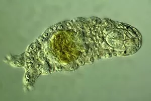
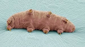
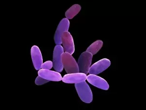
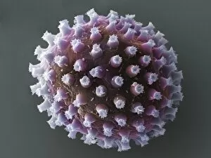
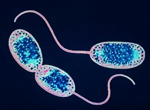
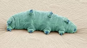
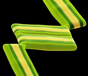
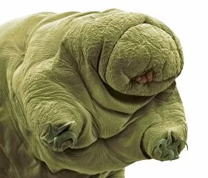
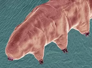
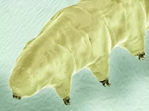
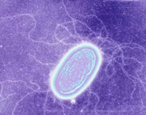
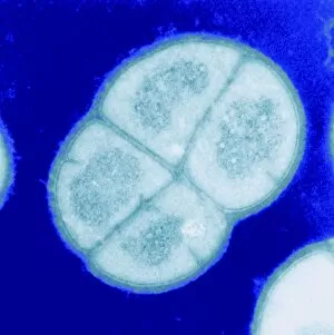
"Exploring the Unseen World: Extremophiles and Their Extraordinary Adaptations" In a world teeming with life, there exists a group of remarkable organisms known as extremophiles. These resilient creatures have evolved to thrive in some of the harshest environments on our planet, pushing the boundaries of what we once thought possible. Take for instance the water bear, also known as Tardigrade. With its pudgy appearance and eight stubby legs, this microscopic creature can withstand extreme conditions that would be fatal to most other organisms. Captured under a light micrograph C016/8581 or SEM C016/9084, these images reveal their incredible resilience. But it's not just water bears that possess such extraordinary abilities. Enter Pyrococcus furiosus archaea and Halobacterium archaea, depicted through stunning artworks like C013/5126. These ancient microbes have adapted to survive in scorching hot volcanic vents and highly saline environments respectively. Even at their earliest stages of life, water bear eggs display an astonishing level of resilience when observed under SEM imaging techniques. The intricate details captured in SEM C016/9085 showcase nature's ability to protect even its tiniest offspring. And let us not forget about Tabellaria diatoms - delicate yet hardy microorganisms found thriving amidst challenging conditions as seen in SEM C016/9599 or 9600. They serve as a testament to how life finds ways to flourish against all odds. Through further exploration using cutting-edge technology like SEM imaging (C016/9083), we uncover more secrets hidden within the microscopic world inhabited by these extremophiles. Each image reveals another glimpse into their fascinating adaptations that allow them to endure hostile surroundings. As we marvel at images like those captured in C016/9086 or 9082 showcasing water bears defying adversity once again, we are reminded of nature's ingenuity.