
Fibril Collection
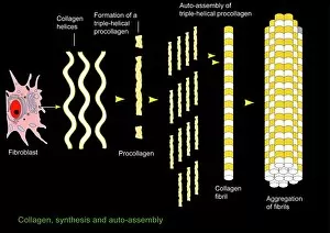
"Fibril: The Intricate Building Blocks of Muscles and More" Collagen synthesis and assembly


For sale as Licensed Images
Choose your image, Select your licence and Download the media
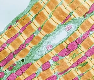
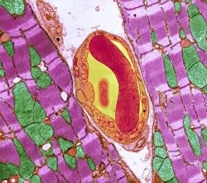
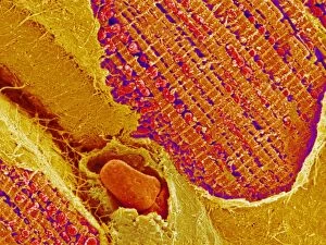
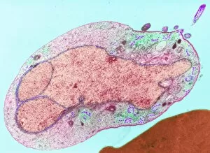
"Fibril: The Intricate Building Blocks of Muscles and More" Collagen synthesis and assembly, artwork: Delve into the fascinating world of fibrils - the intricate building blocks that make up our muscles and beyond. Through collagen synthesis and assembly, these tiny structures play a crucial role in maintaining our body's strength and functionality. Cardiac muscle, TEM: Underneath the microscope, cardiac muscle reveals its mesmerizing beauty. Transmitted Electron Microscopy (TEM) captures the delicate network of fibrils within this vital organ, showcasing their organized arrangement that enables efficient contraction for a healthy heartbeat. Cardiac muscle and capillary, TEM: Zooming even closer with TEM, we witness an extraordinary sight – the intertwining dance between cardiac muscle fibers and capillaries. This intricate relationship ensures optimal oxygen supply to sustain the heart's relentless pumping action. Digital illustration of vertebrate muscle attached to skeleton: Imagine a digital masterpiece illustrating vertebrate muscles connected to their skeletal framework. Within each bundle of muscle fibers lies thousands of thinner yet mighty fibrils working together harmoniously to provide strength, flexibility, and movement. Sugar uptake in muscles diagram: Unlocking one secret behind muscular energy production is understanding sugar uptake within our muscles. A detailed diagram showcases how essential nutrients are absorbed by these remarkable fibrils to fuel our physical activities. Algae cell wall SEM image: Beyond human anatomy lies another realm where fibrils manifest themselves - algae cells. Scanning Electron Microscopy (SEM) unveils an enchanting view of algae cell walls adorned with intricately woven fibrillar patterns that contribute to their structural integrity. Human muscle fibers diagram: Peering into human physiology once again through a comprehensive diagram depicting various types of muscle fibers found in our bodies. These illustrations highlight how different arrangements of fibrils give rise to diverse functions such as endurance or explosive power. Cardiac Muscle SEM image: Another captivating glimpse at cardiac tissue comes from Scanning Electron Microscopy (SEM).