Fibrous Collection (#2)

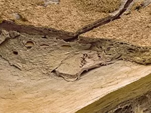
"Fibrous: Unveiling the Intricate Network of Connective Tissues" Liver tissue cirrhosis

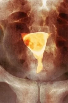
Background imageFibrous Collection: Uterine fibroid, X-ray C014 / 4919

Uterine fibroid, X-ray C014 / 4919
Uterus tumour. Coloured X-ray of the abdomen of a woman with a fibroid (round, centre) in her uterus. A fibroid is a fibrous benign tumour originating from muscular tissue (leimyoma)

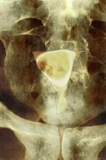
Background imageFibrous Collection: Uterine fibroid, X-ray C014 / 4918

Uterine fibroid, X-ray C014 / 4918
Uterus tumour. Coloured X-ray of the abdomen of a woman with a fibroid (orange, round) in her uterus. A fibroid is a fibrous benign tumour originating from muscular tissue (leimyoma)

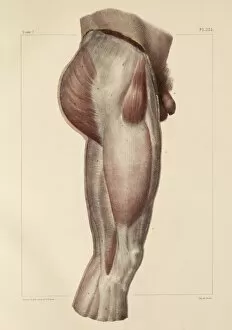
Background imageFibrous Collection: Fascia of the thigh, 1831 artwork

Fascia of the thigh, 1831 artwork
Fascia of the thigh. Lateral view (front of body at right) of the external fascia of the muscles of the thigh. Fascia are fibrous connective tissues that surround muscles and other tissues

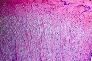
Background imageFibrous Collection: Muscle tissue, light micrograph C014 / 4598

Muscle tissue, light micrograph C014 / 4598
Muscle tissue. Light micrograph of a section through fibrous connective tissue from a longitudinal muscle tendon

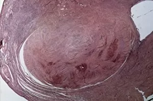
Background imageFibrous Collection: Uterine fibroid, light micrograph C015 / 7101

Uterine fibroid, light micrograph C015 / 7101
Uterine fibroid. Light micrograph of a section through a uterine fibroid (centre). A fibroid is a fibrous benign tumour originating from muscular tissue

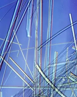
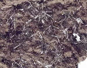
Background imageFibrous Collection: Asbestos fibres, light micrograph

Asbestos fibres, light micrograph
Light micrograph with Normarski Differential Contrast (DIC) illumination of asbestos fibres. Asbestos has been used in the building industry for many years as a result of its heat resistance

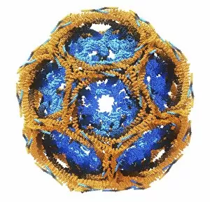
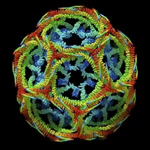
Background imageFibrous Collection: Clathrin lattice, molecular model C015 / 6788

Clathrin lattice, molecular model C015 / 6788
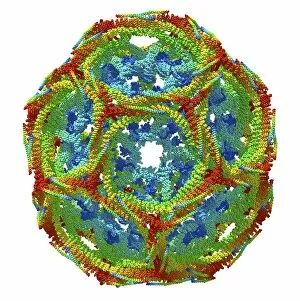
Molecular model of a complete clathrin lattice. The polyhedral protein lattice coats eukaryotic cell membranes (vesicles) and coated pits and appears to be involved in protein secretion

Background imageFibrous Collection: Clathrin lattice, molecular model C015 / 6790

Clathrin lattice, molecular model C015 / 6790
Molecular model of a complete clathrin lattice. The polyhedral protein lattice coats eukaryotic cell membranes (vesicles) and coated pits and appears to be involved in protein secretion

Background imageFibrous Collection: Clathrin lattice, molecular model C015 / 6770

Clathrin lattice, molecular model C015 / 6770
Molecular model of a complete clathrin lattice. The polyhedral protein lattice coats eukaryotic cell membranes (vesicles) and coated pits and appears to be involved in protein secretion

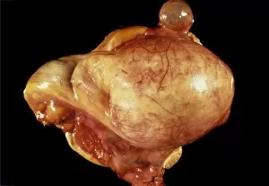
Background imageFibrous Collection: Ovarian fibroid C015 / 6749

Ovarian fibroid C015 / 6749
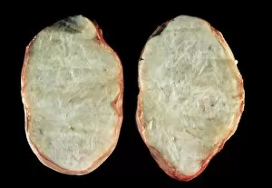
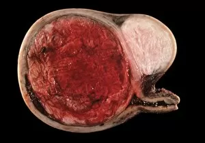
Ovarian fibroid. Gross specimen of a sectioned ovarian fibroid. A fibroid is a fibrous benign tumour originating from muscular tissue

Background imageFibrous Collection: Ovarian fibroid C015 / 6748

Ovarian fibroid C015 / 6748
Ovarian fibroid. Gross specimen of a sectioned ovarian fibroid. A fibroid is a fibrous benign tumour originating from muscular tissue

Background imageFibrous Collection: Uterine fibroid, light micrograph C015 / 6737

Uterine fibroid, light micrograph C015 / 6737
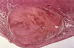
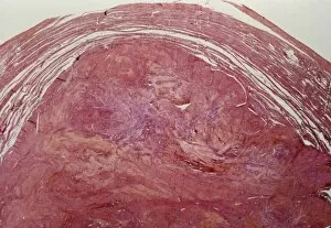
Uterine fibroid. Light micrograph of a section through a uterine fibroid (oval). A fibroid is a fibrous benign tumour originating from muscular tissue

Background imageFibrous Collection: Uterine fibroid C015 / 6416

Uterine fibroid C015 / 6416
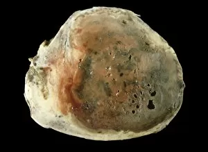
Uterine fibroid. Gross specimen of a sectioned uterine fibroid. A fibroid is a fibrous benign tumour originating from muscular tissue

Background imageFibrous Collection: Uterine fibroid, light micrograph C015 / 6413

Uterine fibroid, light micrograph C015 / 6413
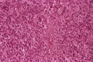
Uterine fibroid. Light micrograph of a section through a uterine fibroid. A fibroid is a fibrous benign tumour originating from muscular tissue

Background imageFibrous Collection: Uterine fibroid C015 / 6415

Uterine fibroid C015 / 6415
Uterine fibroid. Gross specimen of a sectioned uterine fibroid. A fibroid is a fibrous benign tumour originating from muscular tissue

Background imageFibrous Collection: Uterine fibroid, light micrograph C015 / 6411

Uterine fibroid, light micrograph C015 / 6411
Uterine fibroid. Light micrograph of a section through a uterine fibroid (centre). A fibroid is a fibrous benign tumour originating from muscular tissue

Background imageFibrous Collection: Ovarian fibroid C015 / 6414

Ovarian fibroid C015 / 6414
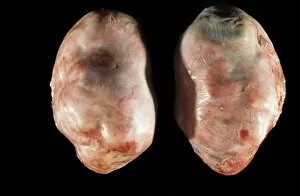
Ovarian fibroid. Gross specimen of an ovarian fibroid. A fibroid is a fibrous benign tumour originating from muscular tissue

Background imageFibrous Collection: Uterine fibroid, light micrograph C015 / 6412

Uterine fibroid, light micrograph C015 / 6412
Uterine fibroid. Light micrograph of a section through a uterine fibroid (oval). A fibroid is a fibrous benign tumour originating from muscular tissue

Background imageFibrous Collection: Uterine fibroid C015 / 6406

Uterine fibroid C015 / 6406
Uterine fibroid. Gross specimen of a uterine fibroid. A fibroid is a fibrous benign tumour originating from muscular tissue

Background imageFibrous Collection: Thomsonite mineral specimen C016 / 4930

Thomsonite mineral specimen C016 / 4930
Thomsonite mineral specimen. Thomsonite consists of hydrated sodium calcium aluminum silicate. It forms tight, fibrous crystals

Background imageFibrous Collection: Benjamin Franklins asbestos purse

Benjamin Franklins asbestos purse. This purse, made from tremolite asbestos, was brought to the UK by 19-year-old future US statesman and scientist Benjamin Franklin (1706-1790) in 1724

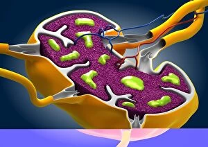
Background imageFibrous Collection: Lymph node, artwork C013 / 4632

Lymph node, artwork C013 / 4632
Lymph node. Computer artwork of a section through a human lymph node. Lymph nodes are kidney-shaped structures located at intervals along lymph vessels (tubes)

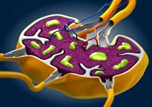
Background imageFibrous Collection: Lymph node, artwork C013 / 4631

Lymph node, artwork C013 / 4631
Lymph node. Computer artwork of a section through a human lymph node. Lymph nodes are kidney-shaped structures located at intervals along lymph vessels (tubes)

Background imageFibrous Collection: Wavellite

Wavellite
Aggregates of honey-coloured wavellite forming radiating needles on slate. Wavellite comprises of (hydrated aluminum phosphate hydroxide). Specimen from Filleigh, Devon

Background imageFibrous Collection: Cerussite

Cerussite
Aggregates of white crystals on globular crystalline baryte. Cerussite comprises of (lead carbonate). Specimen from Frank Mills mine, Christow, Devon

Background imageFibrous Collection: Connellite

Connellite
Deep-blue velvety crust of needles with some red cuprite. Connellite comprises of (hydrated copper sulphate chloride hydroxide). Specimen from Wheal Muttrell, Gwennap, Cornwall

Background imageFibrous Collection: Millerite

Millerite comprises of (nickel sulphide) and is characterized by hair-like fibrous crystals arranged into sprays. Specimen from the collections of the Natural History Museum, London

Background imageFibrous Collection: Jamesonite

Jamesonite comprises of lead iron antimony sulphide, and is characterized by hair-like fibrous crystals. This specimen is from the collections held at the Natural History Museum, London

Background imageFibrous Collection: Tenorite

Tenorite is comprised of (copper oxide). It is found as grey to black metallic crystals and as a by-product of lava flows. Fibrous specimen from the Natural History Museum, London

Background imageFibrous Collection: Liskeardite

Liskeardite
A specimen of the mineral liskeardite from the Marke valley mine, Liskeard, Cornwall. Soft fibrous crystals, forming a pale-green botryoidal crust on a fine-grained matrix of chlorite and quartz

Background imageFibrous Collection: Thomsonite

Thomsonite

Background imageFibrous Collection: Boulangerite

Boulangerite comprises of (lead antimony sulphide) and is characterized by fine, fibrous crystals. This specimen is from the Natural History Museum, London

Background imageFibrous Collection: Asbestos

Asbestos
Geometric asbestos crystals. Asbestos is a dangerous, fibrous amphibole mineral

Background imageFibrous Collection: Asbestos purse

Asbestos purse
Benjamin Franklins Asbestos purse. A crudely plaited purse made from tremolite asbestos. From the Hans Sloane collection. Asbestos is formed of microscopically fibrous crystals

Background imageFibrous Collection: Greater Tussock Sedge (Carex paniculata) tussocks with fibrous rhizomes, in valley fen reserve

Greater Tussock Sedge (Carex paniculata) tussocks with fibrous rhizomes, in valley fen reserve, Hopton Fen, Hopton, Suffolk, England, november

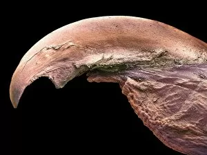
Background imageFibrous Collection: Lovebirds claw, SEM

Lovebirds claw, SEM
Lovebirds claw. Coloured scanning electron micrograph (SEM) of a claw (talon) on the toe of a Lovebird (Agapornis sp.). The claws and beaks of birds are made from keratin, a hard, fibrous protein

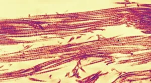
Background imageFibrous Collection: Collagen fibres, TEM

Collagen fibres, TEM
Collagen fibres. Coloured transmission electron micrograph (TEM) of collagen protein fibres. Collagen has a high tensile strength, providing structure and elasticity to skin, tendons

Background imageFibrous Collection: Lactating breast tissue, SEM

Lactating breast tissue, SEM

Background imageFibrous Collection: Retina rod cells, SEM

Retina rod cells, SEM
Retina rod cells. Coloured scanning electron micrograph (SEM) of a freeze-fractured section through a retina, revealing the structure of its photoreceptors

Background imageFibrous Collection: Outer layers of the eye, SEM

Outer layers of the eye, SEM
Outer layers of the eye. Coloured scanning electron micrograph (SEM) of the choroid (top) and sclera (bottom) layers of the eye. The sclera is the white outer layer of the eye

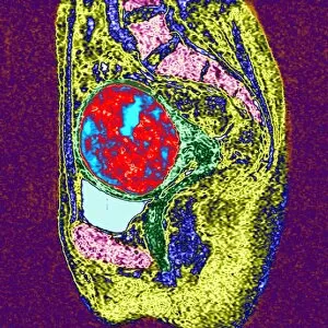
Background imageFibrous Collection: Uterine fibroid, MRI scan

Uterine fibroid, MRI scan

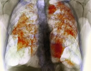
Background imageFibrous Collection: Lung silicosis, X-ray

Lung silicosis, X-ray
Lung silicosis. Coloured X-ray of the lungs of a patient with silicosis, a type of pneumoconiosis. The orange masses in the lungs are areas of fibrous (scarred) tissue and inflammation

Background imageFibrous Collection: Skeletal muscle, SEM

Skeletal muscle, SEM
Skeletal muscle. Scanning electron micrograph (SEM) of a transverse section through skeletal muscle. The many muscle fibres (red) are seen, along with the elastic sheath (epimysium)

Background imageFibrous Collection: Coronary thrombosis, light micrograph

Coronary thrombosis, light micrograph
Coronary thrombosis. Light micrograph of a section through a thrombus (blood clot) in the coronary artery of the heart. The clot is packed full of red blood cells (red)

Background imageFibrous Collection: Fibrosis of the lung, X-ray

Fibrosis of the lung, X-ray
Lung fibrosis. Coloured X-ray of the chest of a patient with pulmonary (lung) fibrosis, or interstitial lung disease. Pulmonary fibrosis is the overgrowth of fibrous scar tissue (dark red)

Background imageFibrous Collection: Alzheimers disease brain cell, TEM

Alzheimers disease brain cell, TEM
Alzheimers disease brain cell. Coloured transmission electron micrograph (TEM) of a neurofibrillary tangle in a nerve cell from the brain of a patient with Alzheimers disease

Background imageFibrous Collection: Polypropylene filaments, SEM

Polypropylene filaments, SEM
Polypropylene filaments. Coloured scanning electron micrograph (SEM) of filaments of the plastic polypropylene, formed by the polymerization of propylene

Background imageFibrous Collection: Filter paper, SEM

Filter paper, SEM
Filter paper. Scanning electron micrograph (SEM) of the cut end of a piece of filter paper showing its many cellulose fibres


For sale as Licensed Images

Choose your image, Select your licence and Download the media

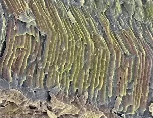
"Fibrous: Unveiling the Intricate Network of Connective Tissues" Liver tissue cirrhosis, light micrograph: A closer look at liver tissue reveals the devastating effects of cirrhosis, where fibrous bands disrupt its normal structure. Tendon, SEM: Under the scanning electron microscope, tendons appear as a dense and resilient fibrous matrix that enables our bodies to move with strength and precision. Collagen synthesis and assembly, artwork: Delicate brushstrokes depict the intricate process of collagen synthesis and assembly – a vital component in forming strong fibrous tissues throughout our body. J. S. Henry Ltd Fireplace and Surrounds: Craftsmanship meets elegance as this stunning fireplace surround showcases its ornate design featuring intricate fibrous patterns carved into high-quality materials. Noyes Bros Advertisement: Step into luxury with Noyes Bros' exquisite collection of furniture upholstered in sumptuous fabrics adorned with subtle yet striking fibrous motifs – an embodiment of refined taste. Plant roots: Fibrous root of groundsel Senecio - Explore nature's engineering marvels as we delve beneath the soil to uncover the complex network of fine fibrous roots supporting plants like groundsel Senecio. Sugar Cane: The tall stalks sway gracefully in fields filled with sugar cane – their tough but flexible fibers hold together this valuable crop that sweetens our lives. Tendon, SEM (again): Another glimpse through the lens reveals tendon fibers interwoven meticulously like a tightly-knit fabric; these resilient structures enable us to perform remarkable feats every day. Page from The Architects Compendium (litho): An architectural masterpiece comes alive on paper as detailed illustrations showcase how innovative designs incorporate both beauty and functionality using various fibrous materials. Page from The Architects Compendium (litho) [repeated].