
Fibula Collection (#3)
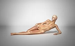
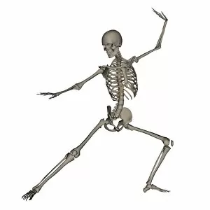
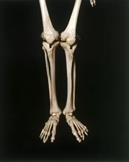
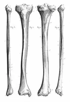
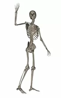
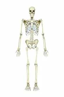
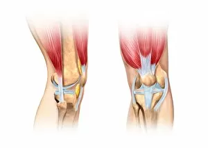
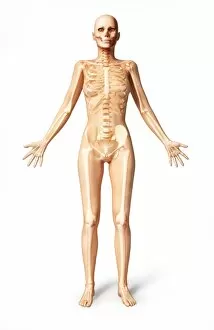
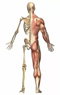
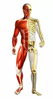
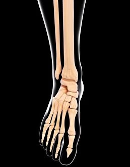
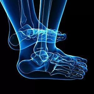
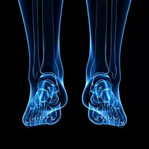
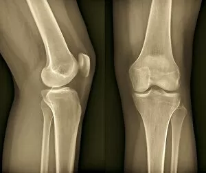
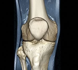
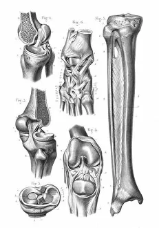
The fibula, a slender bone in the human leg, plays a vital role in supporting the anatomy of the knee joint


For sale as Licensed Images
Choose your image, Select your licence and Download the media
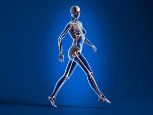
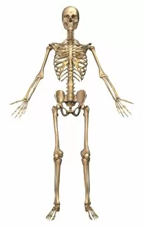
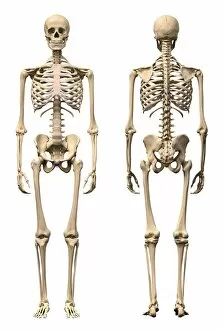
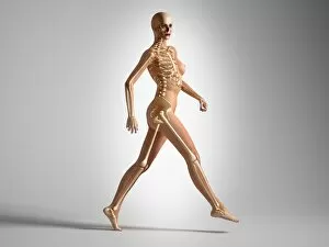
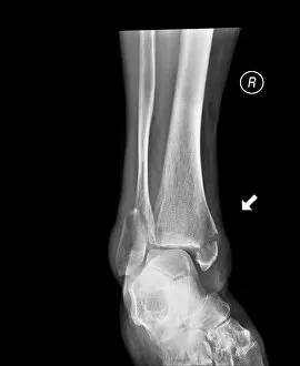
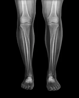
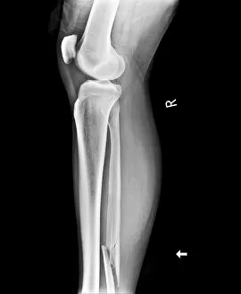
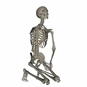
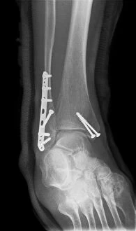
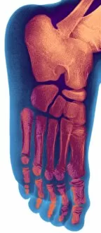
The fibula, a slender bone in the human leg, plays a vital role in supporting the anatomy of the knee joint. Whether it's seen through an X-ray or depicted in artwork, this bone is often associated with various conditions and treatments. In cases where knee joint prosthesis is required, medical professionals rely on their expertise to restore mobility and alleviate pain. X-rays provide valuable insights into damaged knee ligaments, helping doctors determine the best course of action for recovery. Fractured ankles are another common occurrence that can be detected through X-rays. These images showcase the severity of such injuries and guide healthcare providers towards appropriate treatment options. To fully understand the complexity of our lower limbs, diagrams illustrating the bones of the right leg and hip offer a comprehensive view. The fibula stands alongside other crucial components like tibia and femur to ensure stability during movement. However, not all depictions revolve around ailments or injuries. Healthy knees captured through X-rays highlight how well-functioning joints should appear – a testament to proper care and maintenance. Artwork showcasing outer ankle ligaments sheds light on their significance in maintaining balance while inner ankle ligaments receive equal attention as they contribute to overall stability during physical activities. Sometimes extreme measures are necessary for patients suffering from severe knee issues; total knee replacement surgeries become essential interventions. Through x-rays post-surgery, one can observe how these procedures transform lives by restoring functionality. In unfortunate instances where broken legs require stabilization using pins or rods, medical intervention becomes paramount for successful healing processes. These interventions aim at aligning fractured bones accurately so that they can mend properly over time. Elephantiasis may seem unrelated but even this condition has its place when discussing fibulas - x-ray images reveal any potential complications caused by this disease affecting lymphatic vessels within extremities like legs. From anatomy lessons to surgical advancements and artistic representations – each aspect surrounding "fibula" provides unique perspectives on its role in our bodies.