
Filament Collection (#4)
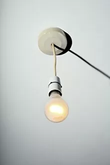
Filament, a word that encompasses a vast array of structures and organisms, holds within it the secrets of life's intricate design


For sale as Licensed Images
Choose your image, Select your licence and Download the media
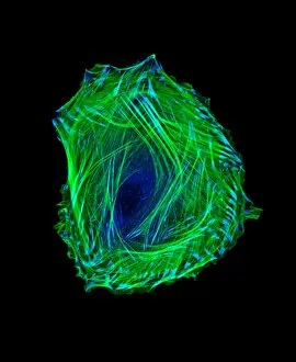
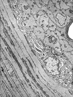
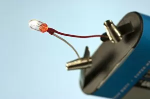
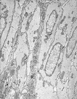
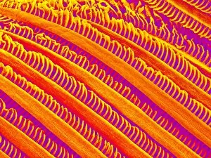
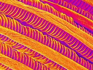
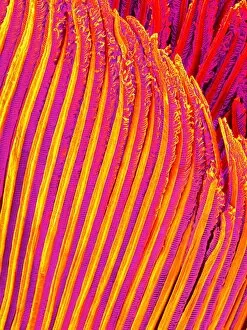
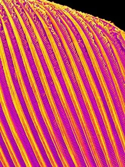
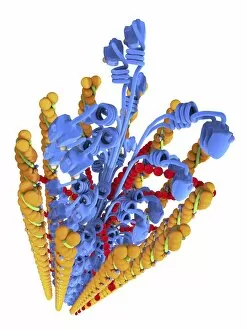
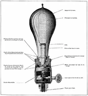
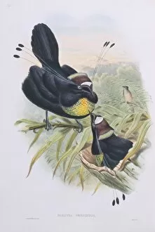
Filament, a word that encompasses a vast array of structures and organisms, holds within it the secrets of life's intricate design. From the budding yeast cell to the HeLa cells captured in stunning light micrographs they can woven into the very fabric of existence. In C017 / 8299, we witness the beauty of HeLa cells under a microscope - delicate strands intertwining like an ethereal dance. These filaments reveal the complexity and interconnectedness of cellular structure. Moving beyond microscopic realms, we encounter geranium anthers through scanning electron microscopy (SEM). The filamentous protrusions on these vibrant flowers serve as nature's invitation for pollinators to partake in their sweet nectar. But filaments extend far beyond plant anatomy; they reach towards celestial wonders too. Solar prominences stretch out like fiery tendrils against the backdrop of our sun's brilliance. These magnificent threads remind us that even in space, filaments connect us to cosmic phenomena. Delving deeper into scientific history, we find Thomas Edison's contribution - an illustration depicting a triode valve. This invention revolutionized technology by controlling electrical currents through filamentary conductors. It paved the way for countless innovations that shape our modern world today. Returning to nature's realm once more, SEM reveals buttercup flowers' intricate details with their captivating filaments delicately embracing pollen grains. Each strand plays its role in ensuring successful reproduction and continuation of life itself. As if painting with science brushes across various disciplines, George Ed captures plate 327 from "The Gleanings of Natural History. " Here he showcases yet another facet: geranium pollen observed under SEM - tiny spheres connected by slender threads forming intricate patterns reminiscent of fine lacework. Filament is not just a word but rather a gateway into understanding life at its core - from cellular biology to astrophysics and everything in between.