
Flatworm Collection (#2)
"Exploring the Fascinating World of Flatworms: From Gastrointestinal Nematodes to Hammerhead Worms" Flatworms, also known as platodes worms


For sale as Licensed Images
Choose your image, Select your licence and Download the media
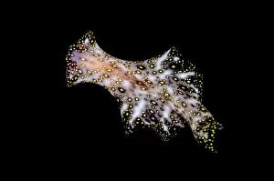
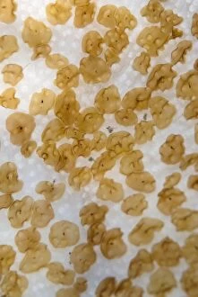
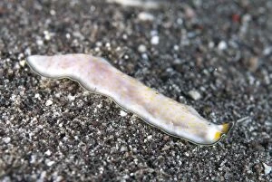
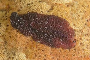
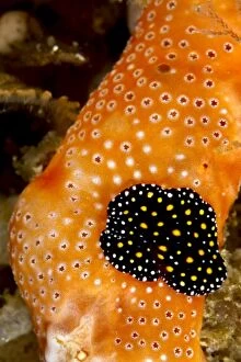
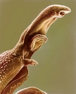
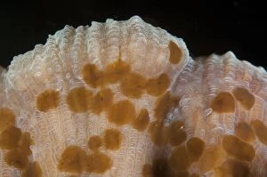
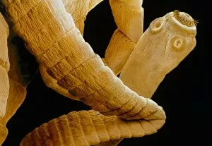
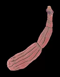
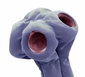
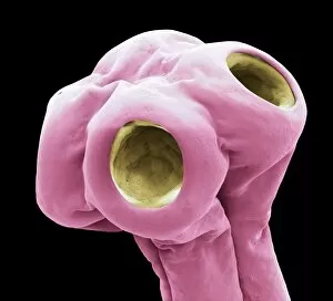
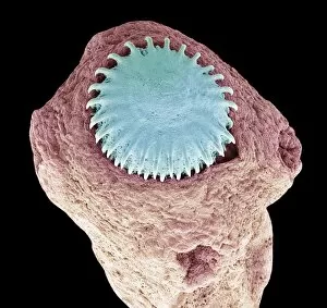
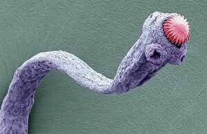
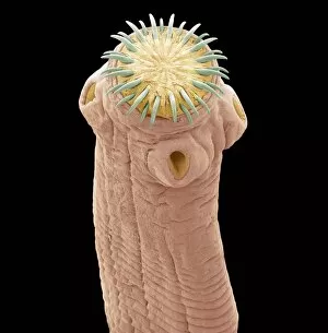
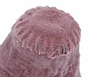
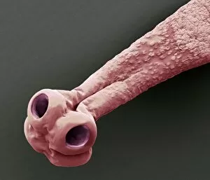
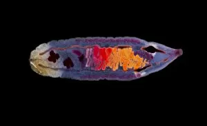
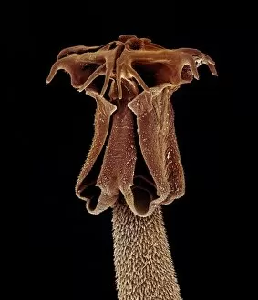
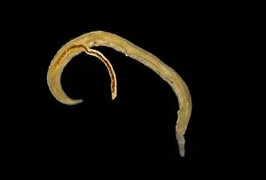
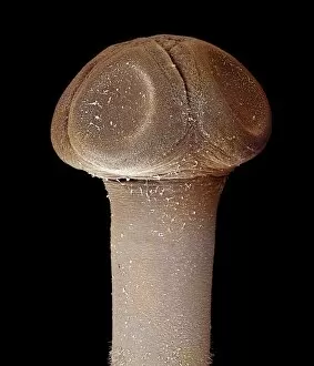
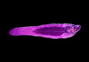
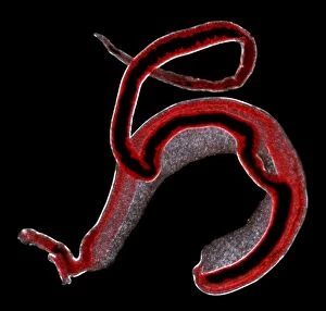
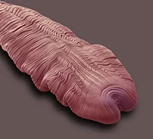
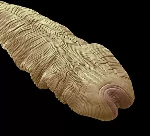
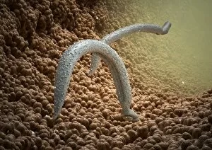
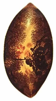
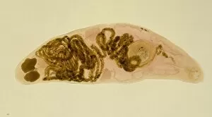
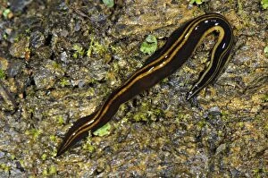
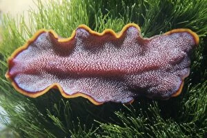
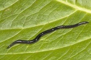
"Exploring the Fascinating World of Flatworms: From Gastrointestinal Nematodes to Hammerhead Worms" Flatworms, also known as platodes worms, are a diverse group of organisms that captivate scientists and nature enthusiasts alike. These intriguing creatures encompass various species with unique characteristics and behaviors. One significant subgroup within flatworms is gastrointestinal nematodes. These microscopic parasites inhabit the digestive tracts of animals, causing diseases and health issues. Their study plays a crucial role in veterinary medicine and public health. Liver flukes are another type that demands attention. They infest the livers of mammals, including humans, leading to severe liver damage if left untreated. Microscope slide preparations allow researchers to observe these intricate creatures up close, unraveling their complex life cycles. Schistosoma spp. , commonly referred to as blood flukes, are yet another captivating member of this phylum. Found in freshwater environments across tropical regions, they cause schistosomiasis—a debilitating disease affecting millions worldwide. Understanding their biology aids in developing effective control strategies. Nature's artistic touch can be witnessed through patterns made in sand by mint-sauce worms (Symsagittifera roscoffensis / Convoluta). These tiny flatworms create mesmerizing trails on sandy shores—an exquisite display of natural beauty. The tapeworm cysticercus offers an astonishing sight when observed under scanning electron microscopy (SEM). This detailed imaging technique reveals the intricacies of its structure—highlighting adaptations for survival within their hosts' intestines. Pictures No. 12479415 and 12479414 showcase the remarkable diversity among flatworm species visually—a testament to nature's creativity at work. Venturing into Sarawak Borneo unveils hammerhead worms (Bipalium sp) gracefully moving along leaves—an enchanting encounter with these elusive creatures amidst lush greenery.