
Fluorescent Light Micrograph Collection

"Unveiling the Intricate World

Background imageFluorescent Light Micrograph Collection: Purkinje nerve cells in the cerebellum

Purkinje nerve cells in the cerebellum
Purkinje cells in the cerebellum. Fluorescent light micrograph of Purkinje cells (green) in the cerebellum of the brain. Purkinje nerve cells have a flask-like body from which numerous highly

Background imageFluorescent Light Micrograph Collection: Kidney tubules in section

Kidney tubules in section
Kidney tubules. Fluorescent light micrograph of a section through kidney tissue showing numerous tubules (black/green). The tubules are seen in cross-section

Background imageFluorescent Light Micrograph Collection: Glial stem cell culture, light micrograph

Glial stem cell culture, light micrograph
Glial stem cell culture. Fluorescent light micrograph of glial stem cells producing the protein NG2 (red) as they mature. These stem cells can differentiate into several types of glial cells

Background imageFluorescent Light Micrograph Collection: Purkinje nerve cells in the cerebellum

Purkinje nerve cells in the cerebellum
Purkinje cells in the cerebellum. Fluorescent light micrograph of Purkinje cells (green) in the cerebellum of the brain. Purkinje nerve cells have a flask-like body from which numerous highly

Background imageFluorescent Light Micrograph Collection: Cerebellum structure, light micrograph

Cerebellum structure, light micrograph
Cerebellum structure. Fluorescent light micrograph of a section through the cerebellum of the brain. The cerebellum comprises three main layers

Background imageFluorescent Light Micrograph Collection: Neural stem cell culture

Neural stem cell culture. Fluorescent light micrograph of a group of neural stem cells (neurosphere) in culture. Neural stem cells are able to differentiate into neurons (nerve cells)

Background imageFluorescent Light Micrograph Collection: Oligodendrocyte nerve cells

Oligodendrocyte nerve cells. Fluorescent light micrograph of human oligodendrocyte nerve cells. Cell nuclei, which contain the cells genetic information, have been dyed blue

Background imageFluorescent Light Micrograph Collection: Astrocyte nerve cell

Astrocyte nerve cell. Fluorescent light micrograph of an astrocyte cell from a human brain. Intermediate filaments (IFs), part of the cells cytoskeleton, have been dyed green

Background imageFluorescent Light Micrograph Collection: Small intestine villi, section

Small intestine villi, section
Villi in the small intestine, fluorescent light micrograph. Villi are finger-like projections from the inner lining of the small intestine that serve to increase the surface area available for

Background imageFluorescent Light Micrograph Collection: Kidney tubules in section

Kidney tubules in section
Kidney tubules. Fluorescent light micrograph of a section through kidney tissue showing numerous tubules (red/green). The tubules are seen in cross-section

Background imageFluorescent Light Micrograph Collection: Neuromuscular synapse, light micrograph

Neuromuscular synapse, light micrograph
Neuromuscular junction. Fluorescent confocal light micrograph of the junction between a nerve cell and a muscle (not seen). The axon of the nerve cell (neuron) has been tagged with a blue dye

Background imageFluorescent Light Micrograph Collection: Retina blood vessel and nerve cells

Retina blood vessel and nerve cells
Retina cells. Fluorescent light micrograph of cells in the retina, the light-sensitive membrane that lines the back of the eyeball

Background imageFluorescent Light Micrograph Collection: Immune response

Immune response. Fluorescent light micrograph of T lymphocytes (red) surrounding glial cells. Glial cells are support cells for neurons (nerve cells)

Background imageFluorescent Light Micrograph Collection: Nerve cell injury response

Nerve cell injury response
Nerve cell response to brain injury. Fluorescent light micrograph of a section through an injured brain. Glial progenitor cells are green and reactive astrocytes

Background imageFluorescent Light Micrograph Collection: Neurosphere culture

Neurosphere culture. Fluorescent light microscope of a group of neural stem cells (neurosphere) in culture. The stem cells are differentiating into neurons (red) and nerve support cells (green)

Background imageFluorescent Light Micrograph Collection: Human epithelial cells

Human epithelial cells. Fluorescent light micrograph of human epithelial cells in culture. The nuclei, which contain the cells genetic information DNA (deoxyribonucleic acid), have been dyed blue

Background imageFluorescent Light Micrograph Collection: Retina blood vessels and nerve cells

Retina blood vessels and nerve cells. Light micrograph showing the blood vessels and nerve cells in the retina of an eye. The retina is the light-sensitive membrane that lines the back of the eyeball

Background imageFluorescent Light Micrograph Collection: Green algae, light micrograph

Green algae, light micrograph
Green algae. Fluorescent light micrograph of the green filamentous alga Klebsormidium sp.. Chlorophyll, the pigment responsible for photosynthesis, is red

Background imageFluorescent Light Micrograph Collection: Soft rush stem, light micrograph

Soft rush stem, light micrograph
Soft rush stem. Fluorescent light micrograph of a cross section through pith from a soft rush (Juncus effusus) stem. The star-shaped aerenchyma tissue (upper frame)

Background imageFluorescent Light Micrograph Collection: Glial stem cell culture, light micrograph

Glial stem cell culture, light micrograph
Glial stem cell culture. Fluorescent light micrograph of glial stem cells producing the proteins NG2 (green) and OLIG2 (oligodendrocyte lineage transcription factor 2, red) as they mature

Background imageFluorescent Light Micrograph Collection: Inner ear hair cells, light micrograph

Inner ear hair cells, light micrograph
Inner ear hair cells. Fluorescent light micrograph of sensory hair cells (blue) from the cochlea of the inner ear. The protein GLAST (glutamate-aspartate transporter) has been labelled red

Background imageFluorescent Light Micrograph Collection: Nerve cell trauma response

Nerve cell trauma response. Fluorescent light micrograph of a section through a spinal cord affected by multiple sclerosis (MS)

Background imageFluorescent Light Micrograph Collection: Neural stem cells in culture

Neural stem cells in culture
Neural stem cell in culture, fluorescent light micrograph. The stem cells have been dyed for nestin (red), an intermediate filament (IF) protein, and the nuclei are dyed blue

Background imageFluorescent Light Micrograph Collection: Brain cells in culture, light micrograph

Brain cells in culture, light micrograph
Brain cells in culture. Fluorescent light micrograph of a microglial cell (upper left) and an oligodendrocyte (centre) from a human brain

Background imageFluorescent Light Micrograph Collection: Nerve cell trauma response

Nerve cell trauma response. Fluorescent light micrograph of a section through a spinal cord affected by multiple sclerosis (MS)


For sale as Licensed Images

Choose your image, Select your licence and Download the media

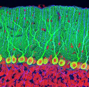
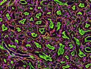
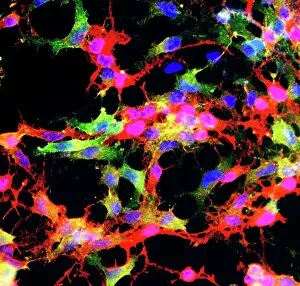
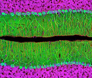
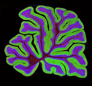
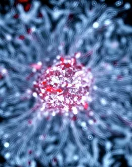
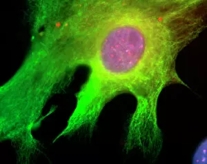
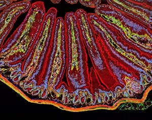
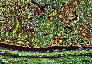
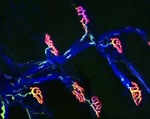
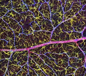
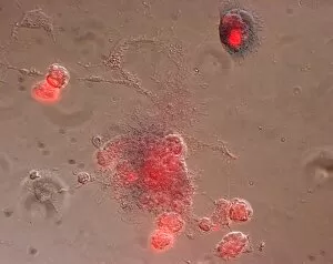
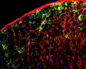
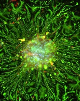
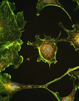
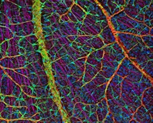
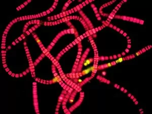
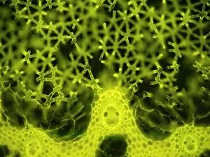
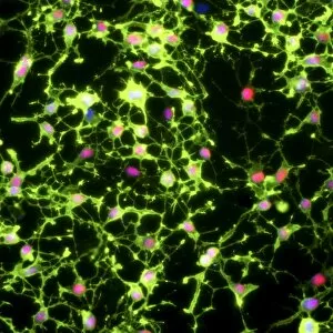
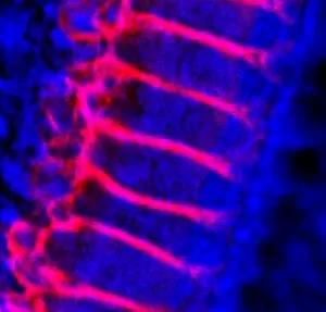
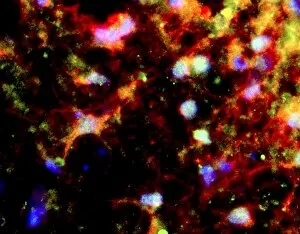
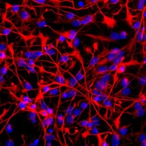
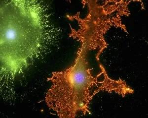
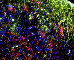
"Unveiling the Intricate World: A Fluorescent Light Micrograph Journey" Step into the mesmerizing realm of cellular structures and witness their vibrant beauty through fluorescent light micrographs. Delve deep into the intricate workings of various tissues and cells, as each image reveals a unique story. Marvel at the Purkinje nerve cells in the cerebellum, with their elaborate branching patterns resembling delicate trees in a forest. These specialized neurons play a crucial role in coordinating movement and maintaining balance. Explore kidney tubules in section, where an array of tiny tubes intricately filter waste products from our blood. Witness how these tubules form an essential part of our renal system, ensuring proper excretion and maintaining fluid balance within our bodies. Behold the glial stem cell culture captured under this illuminating technique. Glial cells are often referred to as "supporting actors" for neurons, providing structural support and nourishment to ensure optimal brain function. Immerse yourself in neural stem cell culture imagery that showcases the remarkable potential for regeneration within our nervous system. These versatile cells hold promise for future therapies aimed at repairing damaged neural tissue caused by injury or disease. Return once more to admire Purkinje nerve cells within the cerebellum structure – their distinctive appearance reminding us of nature's artistry even on a microscopic scale. Witness oligodendrocyte nerve cells, responsible for producing myelin sheaths that insulate neuronal axons. This insulation enables efficient transmission of electrical signals throughout our nervous system – like highways guiding information flow between different regions of our body. Encounter astrocyte nerve cells repeatedly appearing across multiple images - highlighting their significance as star-shaped guardians protecting neurons from harm while also regulating chemical composition around them. Observe kidney tubules again; they serve as vital components involved in filtering waste materials before urine formation – showcasing nature's ingenious design to maintain internal stability continually. Finally, explore small intestine villi in section, where the surface area is magnified to maximize nutrient absorption.