Fluorescent Collection (page 3)

"Unveiling the Vibrant World of Fluorescent Cell Imaging: From Cerebellum Tissue to Dividing Cells" Illuminating the intricate beauty within our brains

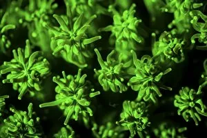
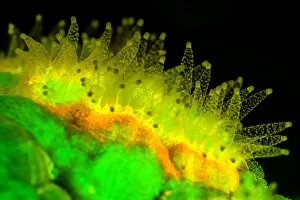
Background imageFluorescent Collection: A coral colony fluoresces as UV light is shown upon it

A coral colony fluoresces as UV light is shown upon it
A coral colony (Galaxea sp.) fluoresces as UV light is shown upon it. Why many marine organisms fluoresce is unknown

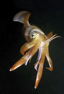
Background imageFluorescent Collection: Bigfin reef squid -Sepioteuthis lessoniana-, Red Sea, Egypt, Africa

Bigfin reef squid -Sepioteuthis lessoniana-, Red Sea, Egypt, Africa

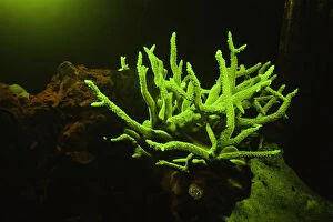
Background imageFluorescent Collection: Fluorescent coral (Acropora sp. ), underwater view

Fluorescent coral (Acropora sp. ), underwater view
This natural fluorescence was captured by using a special ultraviolet light-blocking filter, Rock Islands, Palau, Micronesia

Background imageFluorescent Collection: Picture No. 11091655

Picture No. 11091655
Calcite (red) and Willamite (green) under flourescent light Date:

Background imageFluorescent Collection: Phosphoroscope (1857) by Becquerel, wood engraving, published in 1880

Phosphoroscope (1857) by Becquerel, wood engraving, published in 1880
Phosphoroscope by Alexandre-Edmond Becquerel (French physicist, 1820 - 1891). A phosphoroscope is piece of experimental equipment

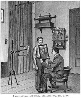
Background imageFluorescent Collection: Examining patients thorax using X-ray tube and fluorescent screen. X-ray tube

Examining patients thorax using X-ray tube and fluorescent screen. X-ray tube (on tripod) is set at reqauired height and patient stands directly in front of it

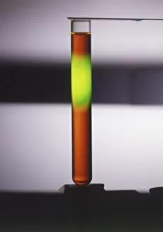
Background imageFluorescent Collection: A test tube contaiing a solution of sodium fluorescein

A test tube contaiing a solution of sodium fluorescein

Background imageFluorescent Collection: Violet-blue fluorite on quartz crystals, fluorescent

Violet-blue fluorite on quartz crystals, fluorescent

Background imageFluorescent Collection: Fluorite crystals on quartz, close up

Fluorite crystals on quartz, close up

Background imageFluorescent Collection: Columnar calcite, fluorescent

Columnar calcite, fluorescent

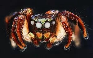
Background imageFluorescent Collection: Jumping spider C018 / 4470

Jumping spider C018 / 4470
Jumping spider. Close-up photograph of a jumping spider specimen preserved in hand sanitiser solution. This specimen (species unknown) was found in the Dominican Republic

Background imageFluorescent Collection: Cotton stem, confocal micrograph C014 / 4636

Cotton stem, confocal micrograph C014 / 4636
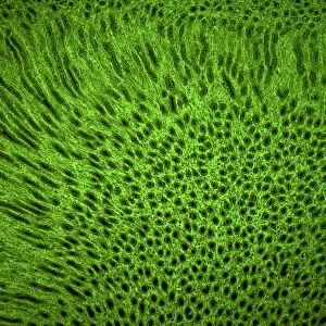
Cotton stem. Laser scanning confocal micrograph (LSCM) of a section through the stem of a cotton (Gossypium sp.) plant

Background imageFluorescent Collection: Stem cell-derived retinal cells

Stem cell-derived retinal cells. Fluorescence light micrograph of retinal pigment epithelium (RPE) cells that have been derived from human embryonic stem cells (HESC)

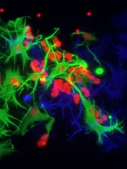
Background imageFluorescent Collection: Astrocyte brain cells, light micrograph

Astrocyte brain cells, light micrograph
Astrocyte brain cells. Fluorescence light micrograph of primary astrocyte cells from the brain of a mouse. Astrocytes have numerous branches of connective tissue that provide support

Background imageFluorescent Collection: Stem cell-derived astrocyte brain cells

Stem cell-derived astrocyte brain cells
Stem cell-derived nerve cells. Fluorescence light micrograph of astrocyte brain cells that have been derived from neural (nerve) stem cells from a mouse

Background imageFluorescent Collection: Crab and coral fluorescing underwater C018 / 9373

Crab and coral fluorescing underwater C018 / 9373
Crab and coral fluorescing underwater under blue light. Some species of underwater animals emit a fluorescent glow. It is thought the glow may attract symbiotic algae

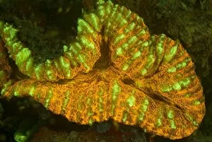
Background imageFluorescent Collection: Lobophyllia coral fluorescing C018 / 9375

Lobophyllia coral fluorescing C018 / 9375
Lobophyllia sp. coral fluorescing under blue light at night. Some species of coral emit a fluorescent glow. It is thought the glow may attract symbiotic algae

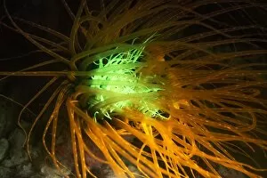
Background imageFluorescent Collection: Cerianthus anemone, fluorescing

Cerianthus anemone, fluorescing
Cerianthus anenome fluorescing under blue or ultraviolet light at night. Photographed in Borneo, Malaysia

Background imageFluorescent Collection: Sea squirt, fluorescing

Sea squirt, fluorescing
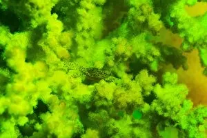
Sea squirt colony (Clavelina sp.) growing in damaged coral. The sea squirt and coral emit a fluorescent light when photographed under ultraviolet or blue light at night

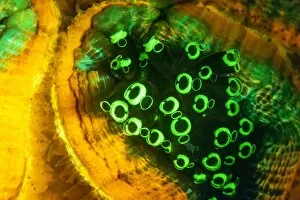
Background imageFluorescent Collection: Favites coral fluorescing

Favites coral fluorescing
Favites sp. coral polyps feeding at night, fluorescing bright orange, yellow and green when illuminated by blue light. Photographed in Borneo, Malaysia

Background imageFluorescent Collection: Squamous epithelium C014 / 4643

Squamous epithelium C014 / 4643
Squamous epithelium. Laser scanning confocal micrograph (LSCM) of a section through stratified squamous epithelial cells. Epithelial tissues line the cavities

Background imageFluorescent Collection: Squamous epithelium, confocal micrograph C014 / 4642

Squamous epithelium, confocal micrograph C014 / 4642
Squamous epithelium. Laser scanning confocal micrograph (LSCM) of a section through stratified squamous epithelial cells. Epithelial tissues line the cavities

Background imageFluorescent Collection: Induced nerve stem cells, micrograph

Induced nerve stem cells, micrograph
Induced nerve stem cells. Fluorescence light micrograph of neural (nerve) stem cells that have been created (induced) from human adult skin fibroblast cells by gene manipulation

Background imageFluorescent Collection: Stem cell-derived nerve cells

Stem cell-derived nerve cells. Fluorescence light micrograph of neural (nerve) stem cells that have been derived from human embryonic stem cells (HESC)

Background imageFluorescent Collection: Lung tissue, confocal micrograph C014 / 4616

Lung tissue, confocal micrograph C014 / 4616
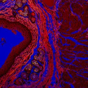
Lung tissue. Laser scanning confocal micrograph (LSCM) of a section through a human lung

Background imageFluorescent Collection: Kidney tissue, confocal micrograph C014 / 4611

Kidney tissue, confocal micrograph C014 / 4611
Kidney tissue. Laser scanning confocal micrograph (LSCM) of a section through a human kidney, showing the cell nuclei (dark)

Background imageFluorescent Collection: Kidney blood vessels, confocal micrograph C014 / 4610

Kidney blood vessels, confocal micrograph C014 / 4610
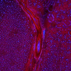
Kidney blood vessels. Laser scanning confocal micrograph (LSCM) of a section through a human kidney, showing a blood vessel (vertical, centre)

Background imageFluorescent Collection: Arabidopsis thaliana embryo, micrograph C014 / 4590

Arabidopsis thaliana embryo, micrograph C014 / 4590
Arabidopsis thaliana embryo. Confocal light micrograph of a section through the embryo of a thale cress (Arabidopsis thaliana) plant

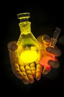
Background imageFluorescent Collection: Fluorescent imaging fluids C015 / 6515

Fluorescent imaging fluids C015 / 6515
Fluorescent imaging fluids. Lab worker holding flasks of fluorescent fluids used in fluorescent imaging for surgical procedures

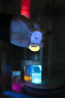
Background imageFluorescent Collection: Fluorescent imaging fluids C015 / 6516

Fluorescent imaging fluids C015 / 6516
Fluorescent imaging fluids. Lab worker holding a vial containing a fluorescent fluid used in fluorescent imaging for surgical procedures

Background imageFluorescent Collection: Children receiving light therapy C015 / 6205

Children receiving light therapy C015 / 6205
Phototherapy. A group of children receiving light therapy (phototherapy). A combination of fluorescent and incandescent lamps are being used

Background imageFluorescent Collection: Municipal worker clearing snow C018 / 2338

Municipal worker clearing snow C018 / 2338
Winter. A municipal worker clearing snow from a courtyard. Photographed in Moscow, Russia

Background imageFluorescent Collection: Thale cress anther and pollen

Thale cress anther and pollen. Confocal laser scanning micrograph of pollen grains (round) on a thale cress (Arabidopsis thaliana) anther
Background imageFluorescent Collection: Fibroblast cell, fluorescent micrograph

Fibroblast cell, fluorescent micrograph
Fibroblast cell. Confocal laser scanning micrograph of a fibroblast cell. The cell nucleus, which contains the cells genetic information, is green

Background imageFluorescent Collection: Plant protoplast, fluorescent micrograph

Plant protoplast, fluorescent micrograph
Protoplast. Confocal laser scanning micrograph of a tobacco plant (Nicotiana tabacum) protoplast. A protoplast is a plant cell that has had its tough outer cell wall removed by chemical treatment

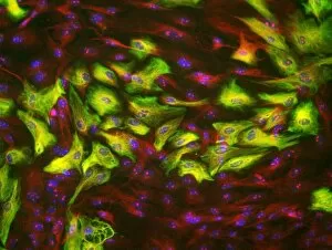
Background imageFluorescent Collection: HeLa cells, light micrograph C013 / 4774

HeLa cells, light micrograph C013 / 4774
HeLa cells. Multi-photon fluorescence light micrograph of a group of cultured HeLa cells, showing the cell nuclei, which contain the cells genetic information (DNA, red)

Background imageFluorescent Collection: HeLa cells, light micrograph C013 / 4773

HeLa cells, light micrograph C013 / 4773
HeLa cells. Multi-photon fluorescence light micrograph of a group of cultured HeLa cells, showing the cell nuclei, which contain the cells genetic information (DNA, blue), and microtubules (pink)

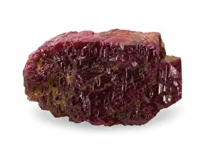
Background imageFluorescent Collection: Natural ruby crystal

Natural ruby crystal
This ruby is a staggering 1, 085 carats. Mined in Burmas Mogok (Myanmar s) mines and bought by the Museum in 1924 from Burma Ruby Mines Ltd. It is one of the largest crystals in the Museum collection

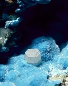
Background imageFluorescent Collection: Susannite on Caledonite

Susannite on Caledonite
4mm crystal of susannite (carbonate) in a mass of caledonite (suplhate). Specimen from the Roughton Gill mine, Caldbeck Fells, Cumbria

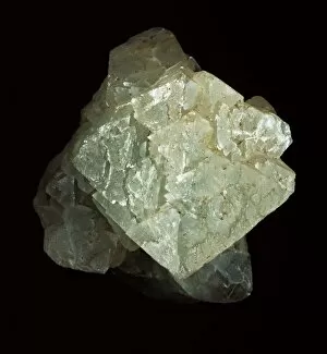
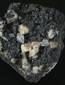
Background imageFluorescent Collection: Fluorite, pale green octahedron

Fluorite, pale green octahedron
With calcite, galena and pyrite. Its properties include fluorescence and cubed crystals. Specimen from Glengowla East mine, Oughterard, Co. Galway, Eire

Background imageFluorescent Collection: Scheelite

Scheelite
Crystals of scheelite (calcium tungstate), an important ore of tungsten from the collections of the Natural History Museum, London

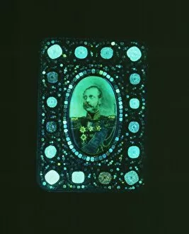
Background imageFluorescent Collection: Diamonds fluorescing

Diamonds fluorescing
Murchison snuff box set with diamonds fluorescing under ultra violet radiation. Gold snuff box set with diamonds ranging from 0.75 to 2.5 carats, presented to Sir R. I. Murchison by Tsar Alexander II

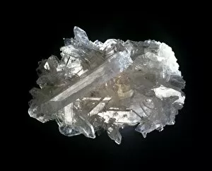
Background imageFluorescent Collection: Celestite

Celestite
A mass of celestite crystals. Celestite is strontium sulphate and is the source of red colours in fireworks

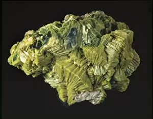
Background imageFluorescent Collection: Autunite

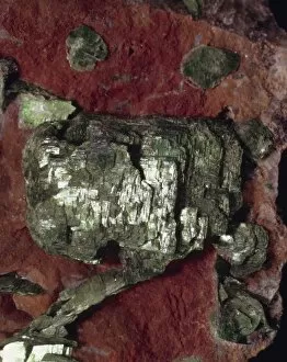
Autunite comprises of (hydrated calcium uranyl phosphate). This is a green, radioactive, highly fluorescent mineral. This specimen is from the Natural History Museum, London

Background imageFluorescent Collection: Franklinite, fluorescing

Franklinite, fluorescing
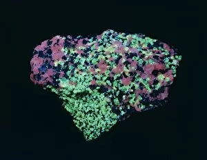
Ore of zinc fluorescing under ultra violet radiation: calcite - pink; willemite - green; zincite - blue; franklinite - black. See image number 388 for white light view

Background imageFluorescent Collection: Willemite

Willemite
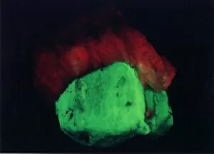
A specimen of willemite (zinc silicate) photographed under UV light

Background imageFluorescent Collection: Torbernite

Torbernite

Background imageFluorescent Collection: Opal doublets

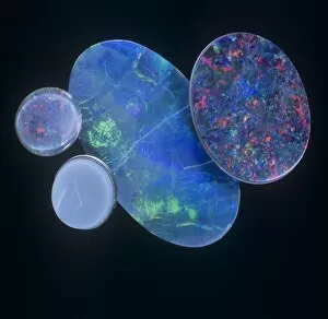
Opal doublets
Opals are not truely crystalline and are therefore mineraloids. They comprise of (hydrated silica glass)


For sale as Licensed Images

Choose your image, Select your licence and Download the media

"Unveiling the Vibrant World of Fluorescent Cell Imaging: From Cerebellum Tissue to Dividing Cells" Illuminating the intricate beauty within our brains, a light micrograph reveals the mesmerizing patterns of cerebellum tissue. Dive into the microscopic realm as glial cells come alive under a confocal light micrograph, showcasing their fluorescent brilliance. HeLa cells take center stage in this captivating light micrograph (C017 / 8299), displaying their striking fluorescence and cellular structure. Witness another stunning view of HeLa cells (C017 / 8298) through a microscope lens, where their vibrant hues reveal hidden secrets within. Journey into the cerebral cortex and marvel at its nerve cells' fluorescent glow, illuminating the complexity of our brain's architecture. Witness the miracle of life itself as cell division unfolds before your eyes in this breathtaking fluorescent micrograph. Returning to explore cerebellum tissue once more, a light micrograph captures its ethereal essence with radiant colors dancing across each frame. The Metropolitan Police launch on the River Thames may seem unrelated but shares an unexpected connection - fluorescence can be found everywhere. Behold nature's perpetual cycle as dividing cells showcase their vivid fluorescence in yet another awe-inspiring image captured by microscopy techniques. Intriguing and visually stunning, these fluorescent hints offer glimpses into various aspects of cellular biology and beyond – from delicate tissues to bustling cityscapes – reminding us that even at microscopic levels, there is beauty all around us waiting to be discovered.