
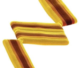
Frustule Collection
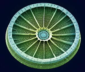
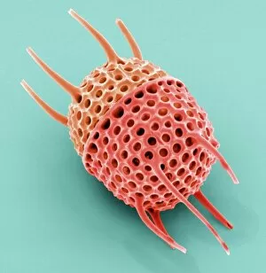
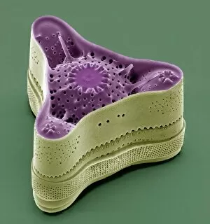
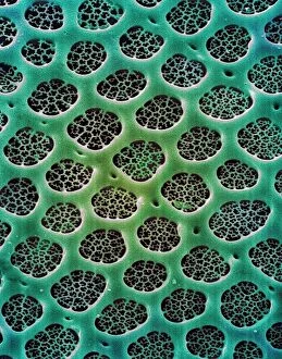
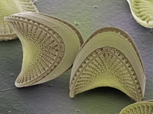
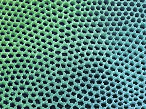
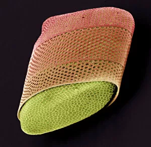
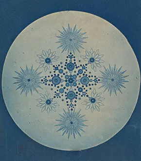
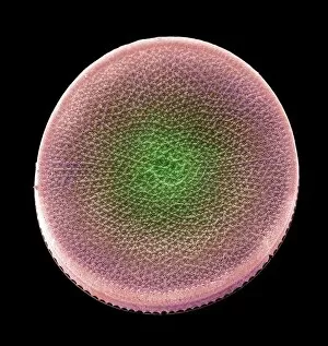
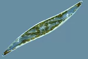
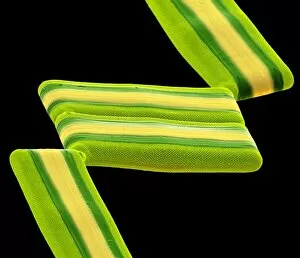
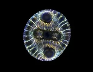
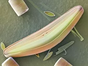
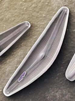
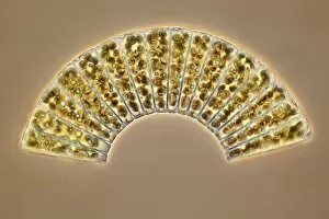
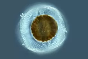
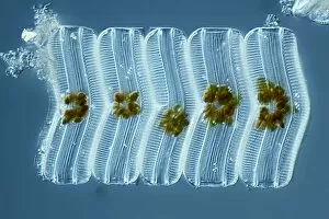
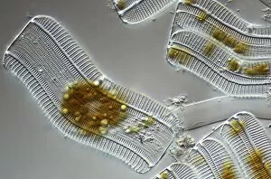
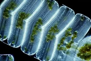
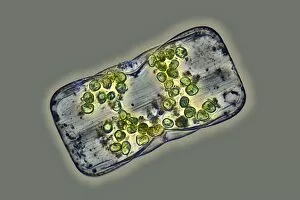
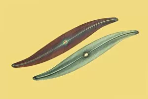
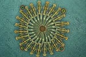
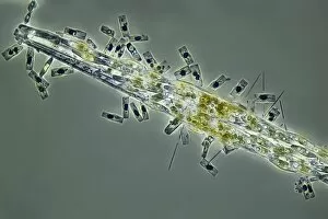
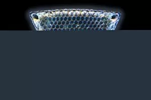
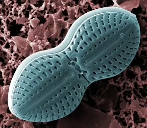
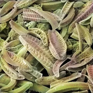
A frustule, the intricate and delicate cell wall of a diatom, is a mesmerizing sight when observed under a scanning electron microscope (SEM


For sale as Licensed Images
Choose your image, Select your licence and Download the media
A frustule, the intricate and delicate cell wall of a diatom, is a mesmerizing sight when observed under a scanning electron microscope (SEM). Diatoms, single-celled algae with silica-based shells, showcase their remarkable beauty through this powerful imaging technique. The SEM allows us to delve into the microscopic world of diatoms, revealing their intricate structures in astonishing detail. The fossilized remains of these ancient organisms can also be examined using SEM, providing valuable insights into Earth's history and evolution. Under the SEM's magnifying lens, we witness the breathtaking complexity of diatom frustules. These tiny wonders display an array of shapes and patterns that vary between species. From elegant spirals to geometric designs resembling ornate stained glass windows - each frustule tells its own unique story. Through SEM imagery, we gain a deeper understanding of how diatoms adapt to their environment. Their exquisitely sculpted shells serve as both protection and support for these microscopic organisms, and is fascinating to observe how different species have evolved distinct frustule structures tailored for survival in various aquatic habitats. The sheer diversity found within diatom communities becomes apparent when examining them under SEM. Countless shapes and sizes come alive on the screen - proof that nature's creativity knows no bounds. This incredible variety showcases the ingenuity behind these minute organisms' ability to thrive across oceans and freshwater bodies worldwide. As we explore further into this hidden realm through SEM technology, our appreciation for the intricacies of life deepens. The stunning beauty captured by these images reminds us that even at such minuscule scales, there exists an awe-inspiring world waiting to be discovered – one where diatoms reign supreme with their captivating frustules as testament to their resilience and artistry.