
General Biology Collection

"Exploring the Microscopic World of General Biology

Background imageGeneral Biology Collection: Malaria plasmodium enlarged under a microscope. Summer-Fall fever. Semi-lunar shapes

Malaria plasmodium enlarged under a microscope. Summer-Fall fever. Semi-lunar shapes. Date of Photograph:1872-1915 ca

Background imageGeneral Biology Collection: Potato starch seen through the lense of a microscope

Potato starch seen through the lense of a microscope Europe. Date of Photograph:2002

Background imageGeneral Biology Collection: Various instruments at Naples Aquarium laboratory

Various instruments at Naples Aquarium laboratory Naples Acquarium. Date of Photograph:1920 - 1930

Background imageGeneral Biology Collection: Laboratory of Naples'Aquarium. In the foreground, two men at work

Laboratory of Naples'Aquarium. In the foreground, two men at work. Naples Acquarium. Date of Photograph:1920 - 1930

Background imageGeneral Biology Collection: Biology. Microscopic enlargment of puccinia graminis microorganisms, wheat parasites

Biology. Microscopic enlargment of puccinia graminis microorganisms, wheat parasites. Date of Photograph:1870-1890 ca

Background imageGeneral Biology Collection: Microscopic study of a cell of nummulite on a slide

Microscopic study of a cell of nummulite on a slide. Date of Photograph:1870-1890 ca

Background imageGeneral Biology Collection: Microscopic enlargement of a micro-organism on a slide

Microscopic enlargement of a micro-organism on a slide. Date of Photograph:1870-1890 ca

Background imageGeneral Biology Collection: Biology. Microscopic enlargement of a micro organism on a slide

Biology. Microscopic enlargement of a micro organism on a slide. Date of Photograph:1870-1890 ca

Background imageGeneral Biology Collection: Planoarcina Ureae enlarged under a microscope

Planoarcina Ureae enlarged under a microscope. Date of Photograph:1914 ca

Background imageGeneral Biology Collection: Microscopic enlargement of Proteus Vulgaris

Microscopic enlargement of Proteus Vulgaris. Date of Photograph:1914 ca

Background imageGeneral Biology Collection: Microscopic enlargement of the cholera vibrio

Microscopic enlargement of the cholera vibrio. Date of Photograph:1872-1915 ca

Background imageGeneral Biology Collection: A bacillus enlarged under a microscope

A bacillus enlarged under a microscope. Date of Photograph:1890-1905 ca

Background imageGeneral Biology Collection: Microscopic enlargement of the Tetanus bacillus

Microscopic enlargement of the Tetanus bacillus. Date of Photograph:1872-1915 ca

Background imageGeneral Biology Collection: Sporiferous tetanus bacillus enlarged under a microscope

Sporiferous tetanus bacillus enlarged under a microscope. Date of Photograph:1872-1915 ca

Background imageGeneral Biology Collection: Streptococcus: bacteria enlarged under a microscope

Streptococcus: bacteria enlarged under a microscope. Date of Photograph:1888-1915 ca

Background imageGeneral Biology Collection: Calliphora Vomitoria (Diptera. Muscidae), from A. Celli, 'Manuale dell'igienista

Calliphora Vomitoria (Diptera. Muscidae), from A. Celli, "Manuale dell'igienista, ad uso di ufficiali sanitari, medici circondariali e provinciali, ingegneri, chimici e veterinari igienisti

Background imageGeneral Biology Collection: Sarcophaga carnaria (Diptera)(Muscidae) from A. Celli, 'Manuale dell'igienista

Sarcophaga carnaria (Diptera)(Muscidae) from A. Celli, "Manuale dell'igienista, ad uso di ufficiali sanitari, medici circondariali e provinciali, ingegneri, chimici e veterinari igienisti

Background imageGeneral Biology Collection: Female Anopheles simplex, magnified under the microscope

Female Anopheles simplex, magnified under the microscope. Date of Photograph:1890-1914 ca

Background imageGeneral Biology Collection: Male Anopheles simplex, magnified under the microscope

Male Anopheles simplex, magnified under the microscope. Date of Photograph:1890-1914 ca

Background imageGeneral Biology Collection: Female Anopheles maculiformis, magnified under the microscope

Female Anopheles maculiformis, magnified under the microscope. Date of Photograph:1890-1914 ca

Background imageGeneral Biology Collection: Microscopic enlargement of Achorion Schoenleinii, parasite fungus of man

Microscopic enlargement of Achorion Schoenleinii, parasite fungus of man. Date of Photograph:1897-1914 ca

Background imageGeneral Biology Collection: Fabulous ringworm fungus on day 16 magnified under the microscope

Fabulous ringworm fungus on day 16 magnified under the microscope. Date of Photograph:1914 ca

Background imageGeneral Biology Collection: Campanularia Dichotoma (Campanularie) Hydromedusa, enlarged under the microscope

Campanularia Dichotoma (Campanularie) Hydromedusa, enlarged under the microscope. Date of Photograph:1914

Background imageGeneral Biology Collection: Culture of Pseudomonas Porrettana in colonies, daisy-shaped magnified under the microscope

Culture of Pseudomonas Porrettana in colonies, daisy-shaped magnified under the microscope. Date of Photograph:01/1905

Background imageGeneral Biology Collection: Gonothyraea Loveni (Polyp-medusae) magnified under the microscope

Gonothyraea Loveni (Polyp-medusae) magnified under the microscope. Date of Photograph:1897

Background imageGeneral Biology Collection: Carbuncle bacillus in a test tube at the tenth day of culture

Carbuncle bacillus in a test tube at the tenth day of culture. Date of Photograph:1872-1915 ca

Background imageGeneral Biology Collection: Separated colonies of Edema bacilli being cultured

Separated colonies of Edema bacilli being cultured. Date of Photograph:1872-1915 ca

Background imageGeneral Biology Collection: Carbuncle bacillus in a test tube at the fifth day of culture

Carbuncle bacillus in a test tube at the fifth day of culture. Date of Photograph:1872-1915 ca

Background imageGeneral Biology Collection: Coli Bacterium in a test tube with a gas development at the third day of culture

Coli Bacterium in a test tube with a gas development at the third day of culture. Date of Photograph:1872-1915 ca

Background imageGeneral Biology Collection: Coli bacterium in a test tube after 7 days of culture

Coli bacterium in a test tube after 7 days of culture. Date of Photograph:1872-1915 ca

Background imageGeneral Biology Collection: Phosphorescent bacillus, enlarged under a microscope, photographed with the same light after an

Phosphorescent bacillus, enlarged under a microscope, photographed with the same light after an hour of air exposure. Date of Photograph:1890-1905 ca

Background imageGeneral Biology Collection: Pseudomonas Porrettana enlarged under a microscope

Pseudomonas Porrettana enlarged under a microscope. Date of Photograph:03/1905

Background imageGeneral Biology Collection: Phosphorescent bacillus with cillia found in an omelette. Enlarged under a microscope

Phosphorescent bacillus with cillia found in an omelette. Enlarged under a microscope. Date of Photograph:02/1903

Background imageGeneral Biology Collection: Pyogenus foetidus bacillus enlarged under a microscope

Pyogenus foetidus bacillus enlarged under a microscope. Date of Photograph:01/1890

Background imageGeneral Biology Collection: Microscopic enlargment of bacillus of Anthrax nested in a kidney glomerule of a Guinea pig

Microscopic enlargment of bacillus of Anthrax nested in a kidney glomerule of a Guinea pig. Date of Photograph:1900-1915 ca

Background imageGeneral Biology Collection: Diptheric bacillus, enlarged under a microscope

Diptheric bacillus, enlarged under a microscope. Date of Photograph:1872-1915 ca

Background imageGeneral Biology Collection: Bacillus Chauvaei, enlarged under a microscope

Bacillus Chauvaei, enlarged under a microscope. Date of Photograph:1872-1915 ca

Background imageGeneral Biology Collection: Sputum with tubercular bacillus, enlarged under a microscope

Sputum with tubercular bacillus, enlarged under a microscope. Date of Photograph:1888-1915 ca

Background imageGeneral Biology Collection: Culture of tubercular bacillus, enlarged under a microscope

Culture of tubercular bacillus, enlarged under a microscope. Date of Photograph:1888

Background imageGeneral Biology Collection: Gonococco: bacteria enlarged under a microscope

Gonococco: bacteria enlarged under a microscope. Date of Photograph:1888-1915 ca

Background imageGeneral Biology Collection: Morphology of bacteria, various aspects in reproduction, microscopic enlargement, plate 5, in W

Morphology of bacteria, various aspects in reproduction, microscopic enlargement, plate 5, in W. Migula "System der Bakterien "Handbuch der Morphologie

Background imageGeneral Biology Collection: Morphology of bacteria, polar granules, lacunae, capsules and pseudo-capsules

Morphology of bacteria, polar granules, lacunae, capsules and pseudo-capsules, microscopic enlargement, plate 47, in W. Migula System der Bakterien "Handbuch der Morphologie

Background imageGeneral Biology Collection: Microscopic enlargment of Saccharomyces Equi

Microscopic enlargment of Saccharomyces Equi. Date of Photograph:1902-1913 ca

Background imageGeneral Biology Collection: Saccharomyces apiculata: microscopic, unicellular fungus, of the Ascomycetes

Saccharomyces apiculata: microscopic, unicellular fungus, of the Ascomycetes, enlarged under a microscope. Date of Photograph:04/1913

Background imageGeneral Biology Collection: Microscopic enlargment of Saccharomyces Cerevisiae

Microscopic enlargment of Saccharomyces Cerevisiae. Date of Photograph:04/1913

Background imageGeneral Biology Collection: Crenothrix polyspora from a Florentine aqueduct magnified under the microscope

Crenothrix polyspora from a Florentine aqueduct magnified under the microscope. Date of Photograph:1913

Background imageGeneral Biology Collection: The bacterium Beggiatoa alba magnified under the microscope

The bacterium Beggiatoa alba magnified under the microscope. Date of Photograph:04/1913

Background imageGeneral Biology Collection: Gelatinous layer on the surface of the waters of the Donzelle spring enlarged under

Gelatinous layer on the surface of the waters of the Donzelle spring enlarged under the microscope, Porretta. Date of Photograph:1905


For sale as Licensed Images

Choose your image, Select your licence and Download the media

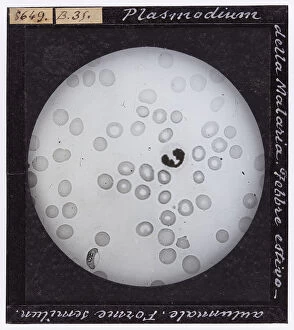
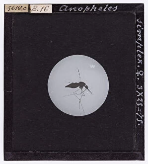
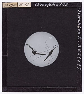
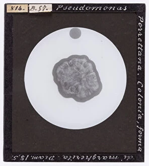
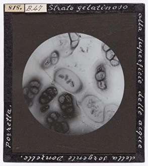
"Exploring the Microscopic World of General Biology: From Enlarged Planoarcina Ureae to Anopheles Maculiformis" Dive into the fascinating realm as we uncover the hidden wonders under the microscope. Witness the intricate details of Planoarcina Ureae, a captivating organism that reveals its beauty when magnified. Marvel at Proteus Vulgaris, with its microscopic enlargement showcasing its unique structure and characteristics. Delve deeper into this microscopic world and encounter the cholera vibrio, an organism notorious for causing disease. Explore a bacillus enlarged under a microscope, revealing its distinct features and potential implications in various biological processes. Discover Tetanus Bacillus in all its glory as it is captured through microscopic enlargement. Witness how this sporiferous bacterium takes on new dimensions when observed closely. Streptococcus bacteria also come to life under our lens, displaying their diverse forms and structures. Venturing beyond bacteria, we explore Calliphora Vomitoria (Diptera. Muscidae) and Sarcophaga carnaria (Diptera) (Muscidae), two intriguing species that captivated scientists over a century ago. These specimens were featured in A. Celli's "Manuale dell'igienista, " offering insights into their significance within hygiene studies during that time. Lastly, meet Anopheles simplex and Anopheles maculiformis – female and male mosquitoes respectively – both meticulously magnified under the microscope. Uncover their distinguishing features as they play crucial roles in transmitting diseases such as malaria. Embark on this visual journey through general biology's microcosmos where every detail holds immense scientific value waiting to be explored.