
Genes Collection (#4)
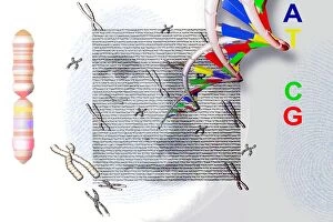
"Unlocking the Secrets: Exploring the Fascinating World of Genes" The X and Y chromosomes: Unraveling the Blueprint of Life


For sale as Licensed Images
Choose your image, Select your licence and Download the media
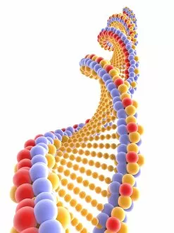
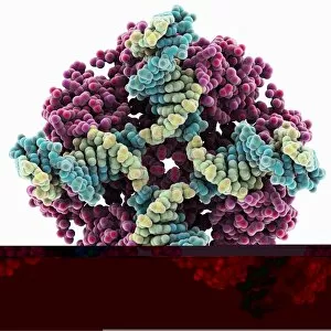
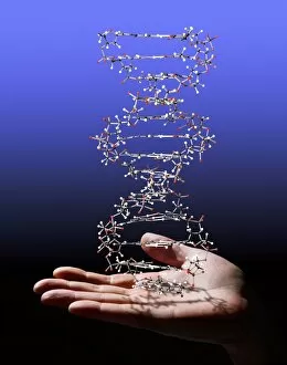
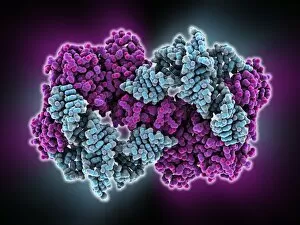
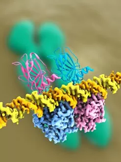
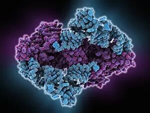
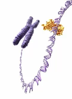
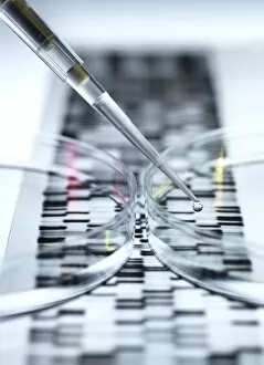
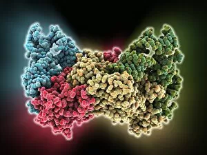
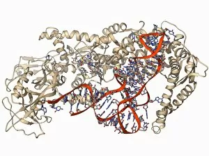
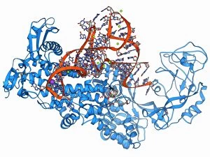
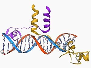
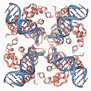
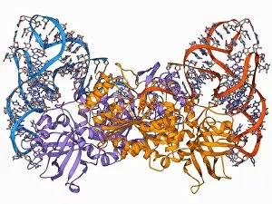
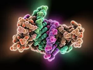
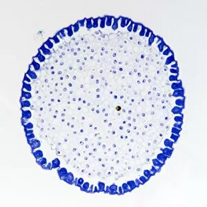
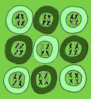
"Unlocking the Secrets: Exploring the Fascinating World of Genes" The X and Y chromosomes: Unraveling the Blueprint of Life. A mesmerizing sight: Leopard's black panther showcases melanistic phase, a result of its genes. DNA molecule: Nature's intricate code for life captured in stunning computer models. Abstract artwork reveals the beauty hidden within the DNA molecule. Gregor Mendel - Pioneering Austrian botanist who laid the foundation for understanding genetic inheritance. Peering into our origins: DNA Double Helix with Autoradiograph offers a glimpse into our genetic makeup. Guinea pigs showcase Mendel's Law through a vibrant poster, highlighting genetic patterns in action. Z-DNA tetramer molecule C015/6557 - Unveiling unique structures within our chromosomes. Chromosomes - The carriers of hereditary information that shape who we are. Delving deep into genetics, this captivating journey takes us from unraveling the mysteries held by X and Y chromosomes to witnessing nature's marvels like a leopard donning its striking black panther coat due to specific genes at play. The awe-inspiring complexity of life is encapsulated in DNA molecules, whether portrayed as computer models or abstract artworks that depict their elegance and intricacy. We pay homage to Gregor Mendel, an Austrian botanist whose groundbreaking work paved the way for understanding how traits are passed down through generations. Through images like DNA Double Helix with Autoradiograph or posters demonstrating Mendel's Laws using guinea pigs as examples, we gain insight into our own origins and witness firsthand how genetics shape every living being on this planet. Zooming further into microscopic wonders, we encounter Z-DNA tetramer molecules and explore fascinating structures found within our very own chromosomes – repositories of invaluable hereditary information.