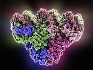
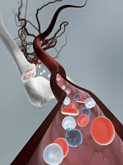
Genetic Collection (page 8)

"Unlocking the Secrets: Exploring the Fascinating World of Genetics" In this captivating journey, we delve into the intricate realm of genetics

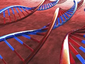
Background imageGenetic Collection: DNA molecules, artwork F006 / 8776

DNA molecules, artwork F006 / 8776
DNA molecules, computer artwork

Background imageGenetic Collection: Genetic engineering, conceptual artwork F006 / 8780

Genetic engineering, conceptual artwork F006 / 8780
Genetic engineering, conceptual computer artwork

Background imageGenetic Collection: DNA molecules, artwork F006 / 8774

DNA molecules, artwork F006 / 8774
DNA molecules, computer artwork

Background imageGenetic Collection: Personal genome sequencing, artwork F006 / 9888

Personal genome sequencing, artwork F006 / 9888
Personal genome sequencing, conceptual computer artwork

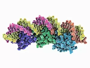
Background imageGenetic Collection: Transcription repressor protein and DNA F006 / 9692

Transcription repressor protein and DNA F006 / 9692
Transcription repressor protein and DNA, molecular model. The repressor protein (green) is binding to a strand of DNA (deoxyribonucleic acid, pink and purple)

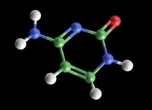
Background imageGenetic Collection: Cytosine molecule, artwork C017 / 7214

Cytosine molecule, artwork C017 / 7214
Cytosine molecule. Computer artwork showing the structure of a molecule of the nucleobase cytosine (2-oxy-4-aminopyrimidine)

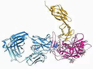
Background imageGenetic Collection: HIV antibody therapy, molecular model F006 / 9622

HIV antibody therapy, molecular model F006 / 9622
HIV antibody therapy. Molecular model of the interaction of the HIV surface protein gp120 (green) as it interacts with a human white blood cell surface protein (CD4)

Background imageGenetic Collection: Aspartyl-tRNA synthetase protein molecule

Aspartyl-tRNA synthetase protein molecule. Molecular model showing the structure of the active site of aspartyl-tRNA synthetase (DARS) from yeast

Background imageGenetic Collection: TATA box-binding protein complex F006 / 9551

TATA box-binding protein complex F006 / 9551
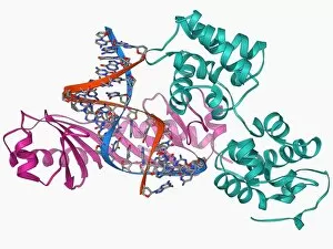
TATA box-binding protein complex. Molecular model showing a yeast TATA box-binding protein (TBP) complexed with a strand of DNA (deoxyribonucleic acid, red and blue) and transcription factor IIA

Background imageGenetic Collection: TATA box-binding protein and DNA F006 / 9550

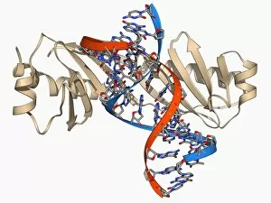
TATA box-binding protein and DNA F006 / 9550
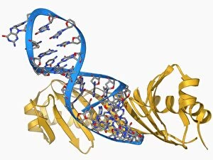
TATA box-binding protein and DNA. Molecular model showing a TATA box-binding protein (TBP) complexed with a strand of DNA (deoxyribonucleic acid, red and blue)

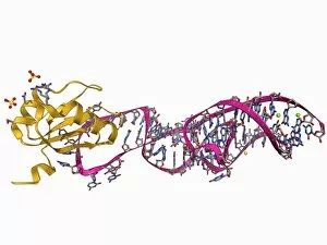
Background imageGenetic Collection: RNA stem-loop motif, molecular model F006 / 9544

RNA stem-loop motif, molecular model F006 / 9544
RNA stem-loop motif. Molecular model of the stem-loop II motif from the SARS (severe acute respiratory syndrome) coronavirus. This RNA (ribonucleic acid) element is a target for antiviral drugs

Background imageGenetic Collection: TATA box-binding protein complex F006 / 9534

TATA box-binding protein complex F006 / 9534
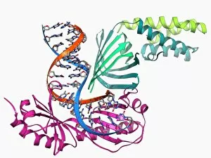
TATA box-binding protein complex. Molecular model showing a yeast TATA box-binding protein (TBP) complexed with a strand of DNA (deoxyribonucleic acid, red and blue) and transcription factor IIB

Background imageGenetic Collection: Transcription activation of IFN-beta gene F006 / 9510

Transcription activation of IFN-beta gene F006 / 9510
Transcription activation of IFN-beta gene. Molecular model of an enhanceosome containing the transcription factors IRF-3, ATF-2 and c-Jun bound to the interferon-beta (IFN-beta)

Background imageGenetic Collection: RNA interference viral suppressor and RNA F006 / 9488

RNA interference viral suppressor and RNA F006 / 9488
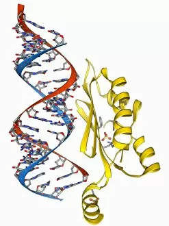
RNA interference viral suppressor and RNA. Molecular model of the p19 protein (yellow) from a Tombusvirus, suppressing a double-stranded, small interfering RNA (siRNA) molecule (red and blue)

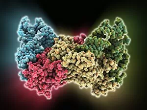
Background imageGenetic Collection: Gene activator protein F006 / 9406

Gene activator protein F006 / 9406
Gene activator protein. Molecular model of catabolite gene activator protein (CAP, yellow) complexed with deoxyribonucleic acid (DNA, red and blue) and RNA polymerase (green and pink)

Background imageGenetic Collection: Valyl-tRNA synthetase molecule F006 / 9342

Valyl-tRNA synthetase molecule F006 / 9342
Valyl-tRNA synthetase protein molecule. Molecular model showing bacterial valyl-tRNA synthetase complexed with valyl tRNA (transfer ribonucleic acid)

Background imageGenetic Collection: DNA Holliday junction complex F006 / 9334

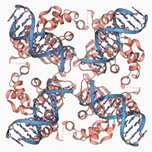
DNA Holliday junction complex F006 / 9334
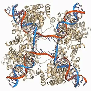
DNA Holliday junction complex. Molecular model of the enzyme FLP recombinase in complex with a Holliday junction between homologous strands of DNA (deoxyribonucleic acid)

Background imageGenetic Collection: Isoleucyl-tRNA synthetase molecule F006 / 9329

Isoleucyl-tRNA synthetase molecule F006 / 9329
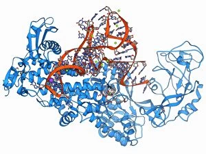
Isoleucyl-tRNA synthetase protein molecule. Molecular model showing bacterial isoleucyl-tRNA synthetase complexed with aspartyl tRNA (transfer ribonucleic acid)

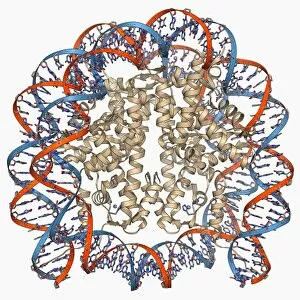
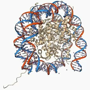
Background imageGenetic Collection: Nucleosome molecule F006 / 9323

Nucleosome molecule F006 / 9323
Nucleosome, molecular model. A nucleosome is a subunit of chromatin, the substance that forms chromosomes. It consists of a short length of DNA (deoxyribonucleic acid)

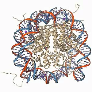
Background imageGenetic Collection: Nucleosome molecule F006 / 9314

Nucleosome molecule F006 / 9314
Nucleosome, molecular model. A nucleosome is a subunit of chromatin, the substance that forms chromosomes. It consists of a short length of DNA (deoxyribonucleic acid)

Background imageGenetic Collection: HP1 molecule C-terminal domain F006 / 9298

HP1 molecule C-terminal domain F006 / 9298
HP1 molecule C-terminal domain. Molecular model showing the structure of the C terminal (shadow chromo) domain of a heterochromatin protein 1 (HP1) molecule from a mouse

Background imageGenetic Collection: Hepatitis D virus ribozyme complex F006 / 9295

Hepatitis D virus ribozyme complex F006 / 9295
Hepatitis D virus ribozyme complex. Molecular model showing an RNA (ribonucleic acid) strand from an Hepatitis delta (Hepatitis D) virus genomic ribozyme, complexed with a ribonucleoprotein

Background imageGenetic Collection: DNA Holliday junction, molecular model F006 / 9285

DNA Holliday junction, molecular model F006 / 9285
DNA Holliday junction. Molecular model of a Holliday junction (centre) between homologous strands of DNA (deoxyribonucleic acid)

Background imageGenetic Collection: Yeast DNA recognition, molecular model F006 / 9282

Yeast DNA recognition, molecular model F006 / 9282
Yeast DNA recognition. Computer model showing a GAL4 transcription activator protein bound to a yeast DNA (deoxyribonucleic acid) molecule (red and blue)

Background imageGenetic Collection: Gene activator protein F006 / 9269

Gene activator protein F006 / 9269
Gene activator protein. Molecular model of catabolite gene activator protein (CAP, pink and green) bound to a molecule of deoxyribonucleic acid (DNA, across top)

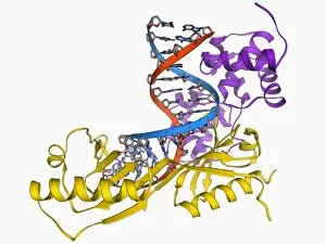
Background imageGenetic Collection: TATA box-binding protein and DNA F006 / 9267

TATA box-binding protein and DNA F006 / 9267
TATA box-binding protein and DNA. Molecular model showing a TATA box-binding protein (TBP) complexed with a strand of DNA (deoxyribonucleic acid, red and blue)

Background imageGenetic Collection: E coli Holliday junction complex F006 / 9261

E coli Holliday junction complex F006 / 9261
E. coli Holliday junction complex. Molecular model of a RuvA protein (red) in complex with a Holliday junction between homologous strands of DNA (deoxyribonucleic acid, blue) from an E

Background imageGenetic Collection: Type II topoisomerase molecule F006 / 9251

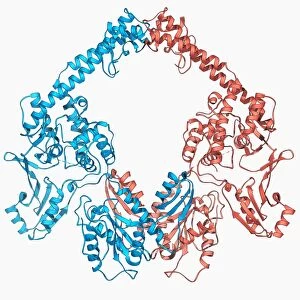
Type II topoisomerase molecule F006 / 9251
Type II topoisomerase, molecular model. The topoisomerase enzymes assist in uncoiling DNA (deoxyribonucleic acid). DNA is usually stored in a supercoiled form

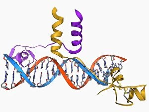
Background imageGenetic Collection: Pit-1 transcription factor bound to DNA F006 / 9242

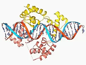
Pit-1 transcription factor bound to DNA F006 / 9242
Pit-1 transcription factor bound to DNA. Molecular model showing pituitary-specific positive transcription factor 1 (Pit-1) (yellow and pink) bound to a strand of DNA (deoxyribonucleic acid)

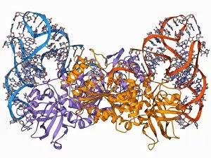
Background imageGenetic Collection: Aspartyl-tRNA synthetase molecule F006 / 9238

Aspartyl-tRNA synthetase molecule F006 / 9238
Aspartyl-tRNA synthetase protein molecule. Molecular model showing bacterial aspartyl-tRNA synthetase complexed with aspartyl tRNA (transfer ribonucleic acid)

Background imageGenetic Collection: Nucleosome molecule F006 / 9235

Nucleosome molecule F006 / 9235
Nucleosome, molecular model. A nucleosome is a subunit of chromatin, the substance that forms chromosomes. It consists of a short length of DNA (deoxyribonucleic acid)

Background imageGenetic Collection: TATA box-binding protein complex F006 / 9230

TATA box-binding protein complex F006 / 9230
TATA box-binding protein complex. Molecular model showing a yeast TATA box-binding protein (TBP) complexed with a strand of DNA (deoxyribonucleic acid, red and blue) and transcription factor IIB

Background imageGenetic Collection: Type I topoisomerase bound to DNA F006 / 9221

Type I topoisomerase bound to DNA F006 / 9221
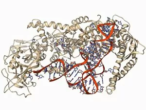
Type I topoisomerase bound to DNA. Molecular model showing a type I topoisomerase molecule (khaki) bound to a strand of DNA (deoxyribonucleic acid, red and blue)

Background imageGenetic Collection: Type I topoisomerase bound to DNA F006 / 9220

Type I topoisomerase bound to DNA F006 / 9220
Type I topoisomerase bound to DNA. Molecular model showing a type I topoisomerase molecule (khaki) bound to a strand of DNA (deoxyribonucleic acid, red and blue)

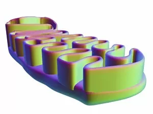
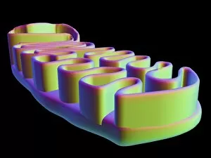
Background imageGenetic Collection: Mitochondrial structure, artwork F006 / 9207

Mitochondrial structure, artwork F006 / 9207
Mitochondrial structure. Computer artwork of a mitochondrion, showing a loop of mitochondrial DNA (deoxyribonucleic acid, mtDNA), the organelles genetic material

Background imageGenetic Collection: Mitochondrial structure, artwork F006 / 9198

Mitochondrial structure, artwork F006 / 9198
Mitochondrial structure. Computer artwork of a mitochondrion, showing a loop of mitochondrial DNA (deoxyribonucleic acid, mtDNA), the organelles genetic material

Background imageGenetic Collection: Zinc finger bound to DNA

Zinc finger bound to DNA. Molecular model showing a zinc finger molecule bound (orange) to a strand of DNA (deoxyribonucleic acid, pink and green)

Background imageGenetic Collection: TATA box-binding protein complex C017 / 7089

TATA box-binding protein complex C017 / 7089
TATA box-binding protein complex. Molecular model showing a TATA box-binding protein (TBP, green) complexed with a strand of DNA (deoxyribonucleic acid, spheres) and transcription factor IIB

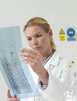
Background imageGenetic Collection: Genetics research F006 / 9088

Genetics research F006 / 9088
MODEL RELEASED. 9Genetics research. Scientist examining a DNA autoradiogram
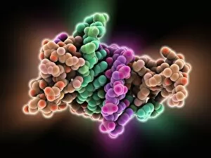
Background imageGenetic Collection: Genetics research F006 / 9087

Genetics research F006 / 9087
MODEL RELEASED. 9Genetics research. Scientist examining a DNA autoradiogram
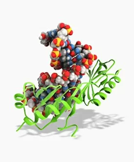
Background imageGenetic Collection: HIV antibody therapy, molecular model C018 / 9193

HIV antibody therapy, molecular model C018 / 9193
HIV antibody therapy. 3D model of the interaction of the HIV surface protein gp120 as it interacts with a human white blood cell surface protein (CD4) and the anti-HIV antibody (17b)

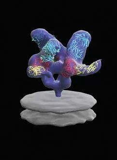
Background imageGenetic Collection: Excisionase complex with DNA

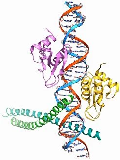
Excisionase complex with DNA. Molecular model of three excisionase proteins (bottom, purple, green and blue) bound to a strand of DNA (top, deoxyribonucleic acid)

Background imageGenetic Collection: Epstein-Barr virus protein and DNA

Epstein-Barr virus protein and DNA. Molecular model of the DNA-binding domain of a viral protein (pink-blue) bound to a lytic gene promoter element (viral strand of DNA, left)

Background imageGenetic Collection: DNA translocase, molecular model

DNA translocase, molecular model
ftsk, , protein, biomolecule, macromolecule, translocase, enzyme, pseudomonas aeruginosa, bacteria, biochemistry, biology, genetics, molecular biology, proteomics, artwork, illustration

Background imageGenetic Collection: RuvBL1 helicase enzyme

RuvBL1 helicase enzyme, molecular model. Helicases are enzymes that carry out several roles, primarily separating the two strands of the DNA (deoxyribonucleic acid) double helix

Background imageGenetic Collection: Metal-binding protein bound to DNA

Metal-binding protein bound to DNA. Molecular model of the bacterial metal-binding protein NikR (bottom) bound to a strand of DNA (top, helical, deoxyribonucleic acid)

Background imageGenetic Collection: Haematopoietic stem cells, artwork

Haematopoietic stem cells, artwork
Haematopoietic stem cells. Cutaway computer artwork showing white blood cells (leucocytes, white, round), red blood cells (erythrocytes, red) and haematopoietic stem cells (HSCs)

Background imageGenetic Collection: Fertilisation, artwork C016 / 7516

Fertilisation, artwork C016 / 7516
Fertilisation. Cutaway computer artwork of a sperm cell (spermatozoa) penetrating an egg (large). The nucleus of the sperm cell will fuse with the egg and its DNA (deoxyribonucleic acid)


For sale as Licensed Images

Choose your image, Select your licence and Download the media

"Unlocking the Secrets: Exploring the Fascinating World of Genetics" In this captivating journey, we delve into the intricate realm of genetics, where computer screens display mesmerizing human genetic sequences. The double-stranded RNA molecule stands as a testament to the complex nature of our genetic makeup. Witness DNA transcription in action through a stunning molecular model, unraveling the process that shapes our very existence. Amidst this exploration, an elegant leopard in its melanistic phase rests gracefully on a log, reminding us of the diversity and beauty found within genes. Computer artwork showcases a beta DNA segment surrounded by spheres, symbolizing both innovation and interconnectedness within our genetic code. The nucleotide base matrix unveils patterns that hold profound significance in understanding hereditary traits. As we peer into abstract images of DNA molecules, we are reminded of their remarkable structure and infinite possibilities they hold for life itself. The intricacies continue with the visualization of nucleosome molecules – tiny structures that play a crucial role in organizing our genetic material. Amidst these wonders lies an HIV reverse transcription enzyme; it serves as a stark reminder of how they can shape not only life but also disease. Yet even amidst challenges, there is hope as scientists tirelessly work to decipher these complexities and find solutions. Ultimately, this captivating journey through various facets of genetics leaves us awestruck by its elegance and complexity. It reminds us that every living being carries within them an extraordinary story written in their DNA – an ancient language connecting all forms of life on Earth.