Genetics Collection (page 6)

"Unlocking the Secrets of Life: Exploring the Fascinating World of Genetics" From the intricate DNA molecule to the X and Y chromosomes

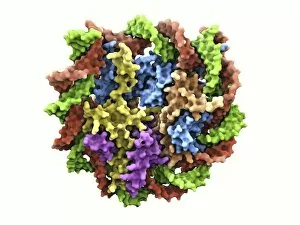
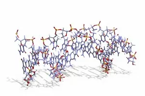
Background imageGenetics Collection: DNA nucleosome, molecular model F007 / 9883

DNA nucleosome, molecular model F007 / 9883
DNA nucleosome. Molecular model of a nucleosome, the fundamental repeating unit used to package DNA (deoxyribonucleic acid) inside cell nuclei
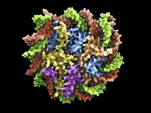
Background imageGenetics Collection: DNA nucleosome, molecular model F007 / 9888

DNA nucleosome, molecular model F007 / 9888
DNA nucleosome. Molecular model of a nucleosome, the fundamental repeating unit used to package DNA (deoxyribonucleic acid) inside cell nuclei

Background imageGenetics Collection: Genetic engineering, conceptual image F007 / 8516

Genetic engineering, conceptual image F007 / 8516
Genetic engineering, conceptual composite image

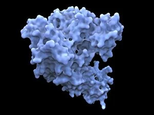
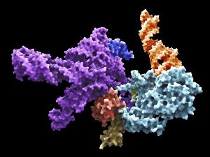
Background imageGenetics Collection: Flap endonuclease protein F007 / 9914

Flap endonuclease protein F007 / 9914
Molecular model of the flap endonuclease protein. This is a class of nucleolytic enzymes that act as both 5 -3 exonucleases

Background imageGenetics Collection: Heat shock factor 70 protein F007 / 9903

Heat shock factor 70 protein F007 / 9903
Molecular model of the Heat Shock Protein 70 (HSP).HSPs are a group of proteins whose levels increase when cells are exposed to raised temperatures or other stress

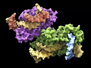
Background imageGenetics Collection: Human 80S ribosome F007 / 9902

Human 80S ribosome F007 / 9902
Ribosomal subunit. Computer model showing the structure of the RNA (ribonucleic acid) molecules in an 80S (large) ribosomal sub-unit. Ribosomes are composed of protein and RNA

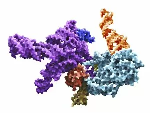
Background imageGenetics Collection: Flap endonuclease protein F007 / 9916

Flap endonuclease protein F007 / 9916
Molecular model of the flap endonuclease protein. This is a class of nucleolytic enzymes that act as both exonucleases and structure-specific endonucleases on specialised DNA structures that occur

Background imageGenetics Collection: Heat shock factor protein F007 / 9892

Heat shock factor protein F007 / 9892
Molecular model of a Heat Shock Protein (HSP).HSPs are a group of proteins whose levels increase when cells are exposed to raised temperatures or other stress

Background imageGenetics Collection: Human 80S ribosome F007 / 9898

Human 80S ribosome F007 / 9898
Ribosomal subunit. Computer model showing the structure of the RNA (ribonucleic acid) molecules in an 80S (large) ribosomal sub-unit. Ribosomes are composed of protein and RNA

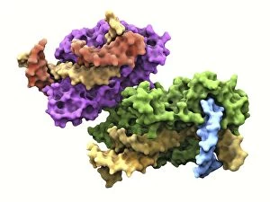
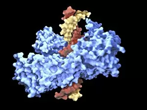
Background imageGenetics Collection: Type I topoisomerase protein bound to DNA F007 / 9893

Type I topoisomerase protein bound to DNA F007 / 9893
Type I topoisomerase bound to DNA. Molecular model showing a type I topoisomerase molecule (blue) bound to a strand of DNA (deoxyribonucleic acid, yellow and red)

Background imageGenetics Collection: DNA sequence, artwork F008 / 3416

DNA sequence, artwork F008 / 3416
DNA (deoxyribonucleic acid) sequence, computer artwork

Background imageGenetics Collection: DNA molecule, artwork F008 / 3371

DNA molecule, artwork F008 / 3371
DNA molecule and chromosome, artwork

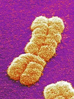
Background imageGenetics Collection: Human chromosome

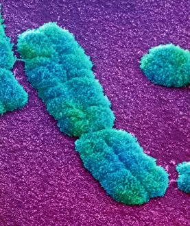
Human chromosome. Coloured scanning electron micrograph (SEM) of a human chromosome. Chromosomes occur in the nucleus of every cell in the body

Background imageGenetics Collection: Genetics research F008 / 3192

Genetics research F008 / 3192
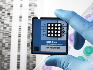
Genetics research

Background imageGenetics Collection: DNA sequence, artwork F008 / 3420

DNA sequence, artwork F008 / 3420
DNA (deoxyribonucleic acid) sequence, computer artwork

Background imageGenetics Collection: Genetics research, conceptual image F008 / 2122

Genetics research, conceptual image F008 / 2122
Genetics research, conceptual image

Background imageGenetics Collection: DNA molecules, artwork F008 / 3372

DNA molecules, artwork F008 / 3372
DNA molecules, artwork

Background imageGenetics Collection: DNA sequence, artwork F008 / 3422

DNA sequence, artwork F008 / 3422
DNA (deoxyribonucleic acid) sequence, computer artwork

Background imageGenetics Collection: Giant chromosomes, SEM P657 / 0034

Giant chromosomes, SEM P657 / 0034
Giant chromosomes. Coloured scanning electron micrograph (SEM) of giant (polytene) chromosomes from a fruit fly (Drosophila busckii)

Background imageGenetics Collection: Genetics research F008 / 3193

Genetics research F008 / 3193
Genetics research

Background imageGenetics Collection: Genetic modification, conceptual image F008 / 2056

Genetic modification, conceptual image F008 / 2056
Genetically modified plant, conceptual image

Background imageGenetics Collection: DNA sequence, artwork F008 / 3417

DNA sequence, artwork F008 / 3417
DNA (deoxyribonucleic acid) sequence, computer artwork

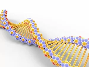
Background imageGenetics Collection: DNA molecule, artwork F008 / 2034

DNA molecule, artwork F008 / 2034
DNA molecule, computer artwork

Background imageGenetics Collection: DNA molecule, artwork F008 / 3370

DNA molecule, artwork F008 / 3370
DNA molecule, artwork

Background imageGenetics Collection: DNA sequence, artwork F008 / 3426

DNA sequence, artwork F008 / 3426
DNA (deoxyribonucleic acid) sequence, computer artwork

Background imageGenetics Collection: DNA molecules, artwork F008 / 3264

DNA molecules, artwork F008 / 3264
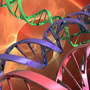
Deoxyribonucleic acid (DNA) molecules, computer artwork

Background imageGenetics Collection: Human chromosome pair, SEM

Human chromosome pair, SEM
Human chromosome. Coloured scanning electron micrograph (SEM) of a human chromosome as a pair of identical copies called chromatids. These form as part of chromosome replication during cell division

Background imageGenetics Collection: DNA sequence, artwork F008 / 3293

DNA sequence, artwork F008 / 3293
DNA sequence, computer artwork

Background imageGenetics Collection: DNA sequence, artwork F008 / 3415

DNA sequence, artwork F008 / 3415
DNA (deoxyribonucleic acid) sequence, computer artwork

Background imageGenetics Collection: DNA sequence, artwork F008 / 3423

DNA sequence, artwork F008 / 3423
DNA (deoxyribonucleic acid) sequence, computer artwork

Background imageGenetics Collection: DNA sequence, artwork F008 / 3418

DNA sequence, artwork F008 / 3418
DNA (deoxyribonucleic acid) sequence, computer artwork

Background imageGenetics Collection: DNA sequence, artwork F008 / 3424

DNA sequence, artwork F008 / 3424
DNA (deoxyribonucleic acid) sequence, computer artwork

Background imageGenetics Collection: DNA sequence, artwork F008 / 3421

DNA sequence, artwork F008 / 3421
DNA (deoxyribonucleic acid) sequence, computer artwork

Background imageGenetics Collection: Genetics research, conceptual image F008 / 2093

Genetics research, conceptual image F008 / 2093
Genetics research, conceptual image

Background imageGenetics Collection: Human genome, conceptual artwork F008 / 3292

Human genome, conceptual artwork F008 / 3292
Human genome, conceptual computer artwork

Background imageGenetics Collection: DNA molecule, artwork F008 / 2036

DNA molecule, artwork F008 / 2036
DNA molecule, computer artwork

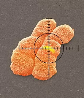
Background imageGenetics Collection: Targeted gene on a chromosome, SEM

Targeted gene on a chromosome, SEM
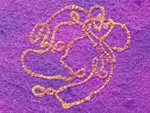
Targeted gene on a human chromosome. Composite coloured scanning electron micrograph (SEM) of a targeted gene (yellow) on one arm of a human chromosome

Background imageGenetics Collection: Genetics research F008 / 3195

Genetics research F008 / 3195
Genetics research

Background imageGenetics Collection: DNA sequence, artwork F008 / 3427

DNA sequence, artwork F008 / 3427
DNA (deoxyribonucleic acid) sequence, computer artwork

Background imageGenetics Collection: DNA molecule F008 / 3657

DNA molecule F008 / 3657
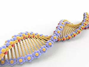
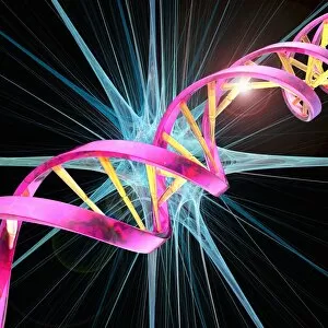
DNA molecule. Computer artwork of the structure of deoxyribonucleic acid (DNA) with a double helix in the background. DNA is composed of two strands twisted into a double helix

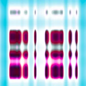
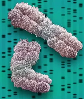
Background imageGenetics Collection: Chromosomes, SEM

Chromosomes, SEM
Chromosomes. Coloured scanning electron micrograph (SEM) of two chromosomes. The SEM is overlaid on a DNA autoradiogram background

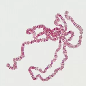
Background imageGenetics Collection: Giant chromosomes, light micrograph P657 / 0038

Giant chromosomes, light micrograph P657 / 0038
Giant chromosomes. Light micrograph of giant (polytene) chromosomes from a fruit fly (Drosophila busckii). These chromosomes are formed from repeated rounds of DNA (deoxyribonucleic acid)

Background imageGenetics Collection: DNA molecules, artwork F008 / 3266

DNA molecules, artwork F008 / 3266
Deoxyribonucleic acid (DNA) molecules, computer artwork

Background imageGenetics Collection: Genetics research F008 / 3191

Genetics research F008 / 3191
Genetics research

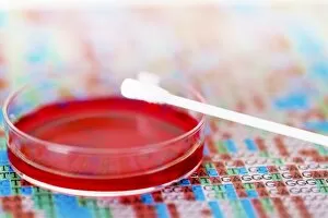
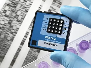
Background imageGenetics Collection: DNA sample tube F008 / 2088

DNA sample tube F008 / 2088
DNA sample tube

Background imageGenetics Collection: DNA sequence, artwork F008 / 3419

DNA sequence, artwork F008 / 3419
DNA (deoxyribonucleic acid) sequence, computer artwork

Background imageGenetics Collection: Genetic research F008 / 2087

Genetic research F008 / 2087
Genetic research

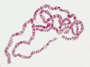
Background imageGenetics Collection: Giant chromosomes, light micrograph P657 / 0037

Giant chromosomes, light micrograph P657 / 0037
Giant chromosomes. Light micrograph of giant (polytene) chromosomes from a fruit fly (Drosophila busckii). These chromosomes are formed from repeated rounds of DNA (deoxyribonucleic acid)


For sale as Licensed Images

Choose your image, Select your licence and Download the media

"Unlocking the Secrets of Life: Exploring the Fascinating World of Genetics" From the intricate DNA molecule to the X and Y chromosomes, a captivating field that unravels the blueprint of life. As we peer into a computer screen displaying a human genetic sequence, we witness the complexity encoded within our very cells. The double-stranded RNA molecule serves as a messenger, carrying vital information for DNA transcription. Molecular models illustrate how this process shapes our traits and characteristics. It was through their groundbreaking work that Watson and Crick discovered the structure of DNA, forever changing our understanding of genetics. Richard Dawkins, an esteemed British science writer, has played an influential role in popularizing genetics among masses. His insightful writings have shed light on evolutionary biology and its connection to our genetic makeup. Intriguingly captured by scanning electron microscopy (SEM), an embryonic stem cell alongside a needle reminds us of the immense potential held within these tiny building blocks. Mitosis comes alive under a light micrograph, showcasing how cells divide and multiply with precision. Computer artwork depicting beta DNA segments interlaced with spheres hints at ongoing research pushing boundaries in genetic engineering. The nucleotide base matrix acts as a foundation for decoding genetic information - each letter representing crucial instructions embedded within our genes. Genetics holds endless possibilities - from unraveling hereditary diseases to designing personalized medicine based on individual genomes. With every discovery made in this ever-evolving field, humanity inches closer towards harnessing nature's codebook for better health and understanding ourselves more deeply than ever before.