
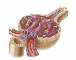
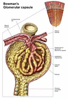
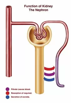
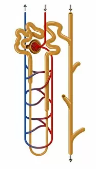
Glomerulus Collection
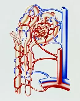
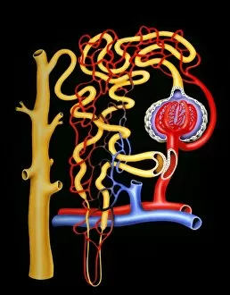
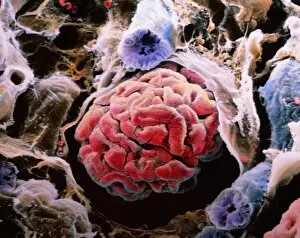
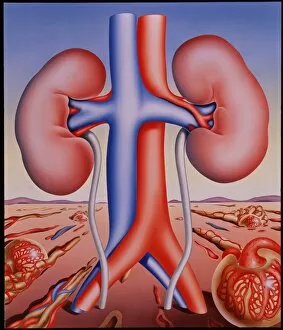
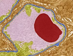
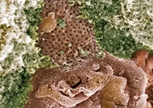
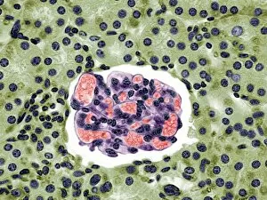
The glomerulus is a vital component of the kidney, responsible for filtering waste and excess fluids from our blood


For sale as Licensed Images
Choose your image, Select your licence and Download the media
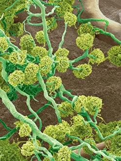
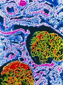
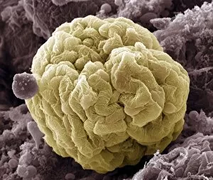
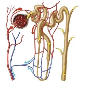
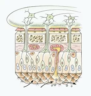
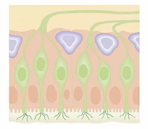
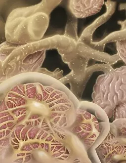
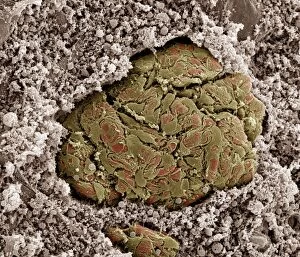
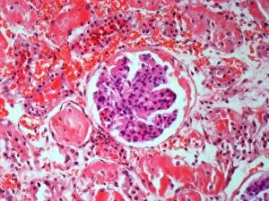
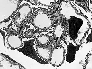
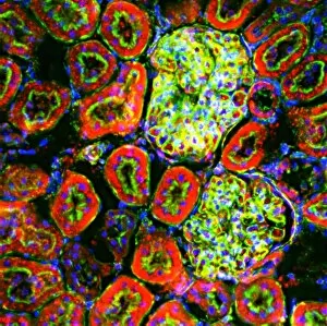
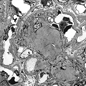
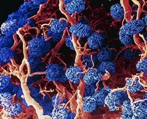
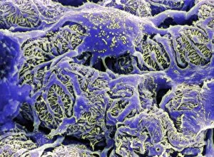
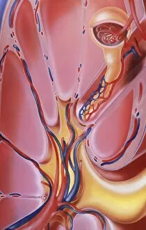
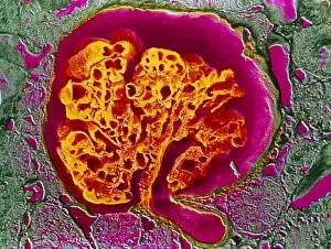
The glomerulus is a vital component of the kidney, responsible for filtering waste and excess fluids from our blood. In this captivating SEM image, we get an up-close look at the intricate network of kidney glomeruli. The delicate structures resemble tiny clusters of grapes, each one playing a crucial role in maintaining our body's fluid balance. Another stunning SEM image showcases the complexity and beauty of the kidney glomerulus. These interconnected spheres are like microscopic sieves, selectively allowing certain substances to pass through while retaining essential molecules within our bloodstream. Zooming out slightly, we observe a mesmerizing view of kidney blood vessels surrounding the glomeruli in yet another striking SEM image. This intricate web ensures efficient transport of filtered substances throughout our body while maintaining proper circulation. Moving away from electron microscopy, a light micrograph provides us with a different perspective on the kidney glomeruli. The vibrant colors highlight their unique structure and emphasize their importance as functional units within our excretory system. Stepping back even further, an artwork illustrates the nephron structure – where these remarkable glomeruli reside – giving us insight into how they fit into the larger framework of renal function. Each nephron acts as an individual processing unit that contributes to overall waste elimination in our kidneys. Shifting gears momentarily, we delve into another fascinating topic: olfactory receptors in nasal cavities. A cross-section biomedical illustration reveals these specialized cells responsible for detecting scents and triggering our sense of smell. While unrelated to kidneys or glomeruli directly, it serves as a reminder that various systems work together harmoniously within our bodies. Returning to focus on renal anatomy once more, Picture No. 11675578 captures an exquisite close-up view of nephron details under high magnification; every minuscule component is meticulously depicted here for scientific examination and appreciation. Lastly but not leastly (if such word exists), Picture No. 11675643 presents a cross-sectional illustration of the human olfactory system.