Gullet Collection (#2)

"The Gullet: A Journey Through Time and Culture" Step into the historical charm of Stafford Street, Birmingham

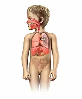
Background imageGullet Collection: Anatomy of a childs full respiratory system

Anatomy of a childs full respiratory system, including mouth and nasal cross section. Other internal organs appear in half tone

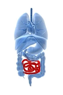
Background imageGullet Collection: X-ray image of internal organs with small intestine highlighted in red

X-ray image of internal organs with small intestine highlighted in red

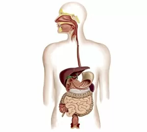
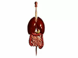
Background imageGullet Collection: Anatomy of human digestive system

Anatomy of human digestive system

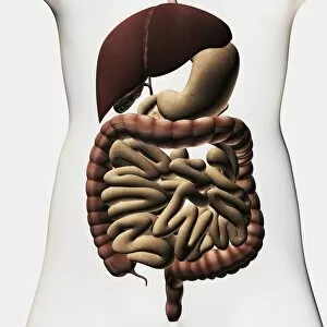
Background imageGullet Collection: Medical illustration showing the human digestive system

Medical illustration showing the human digestive system: liver, stomach, large intestine, small intestine

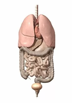
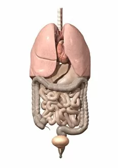
Background imageGullet Collection: Front view of human digestive system with lungs

Front view of human digestive system with lungs

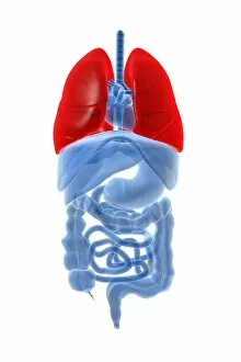
Background imageGullet Collection: X-ray image of internal organs with lungs highlighted in red

X-ray image of internal organs with lungs highlighted in red

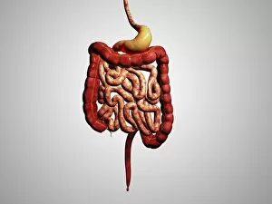
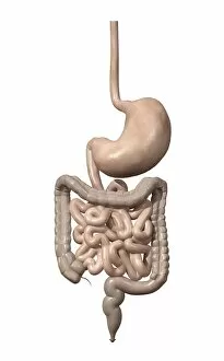
Background imageGullet Collection: Front view of human digestive system

Front view of human digestive system

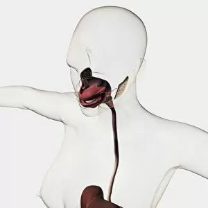
Background imageGullet Collection: Medical illustration of the human digestive system

Medical illustration of the human digestive system; salivary glands, esophagus, oral cavity

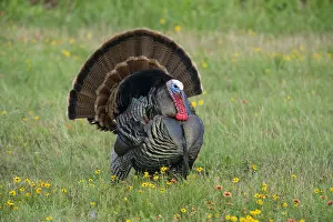
Background imageGullet Collection: USA, Texas, Aransas National Wildlife Refuge, Wild Turkey (Meleagris gallopavo)

USA, Texas, Aransas National Wildlife Refuge, Wild Turkey (Meleagris gallopavo)

Background imageGullet Collection: Illustration of esophagus

Illustration of esophagus
Medicine: Human anatomy, esophagus. Drawing

Background imageGullet Collection: Human intestines, artwork

Human intestines, artwork
Human intestines. Artwork of a human torso with the intestines highlighted in colour. They consist of the small intestine (pink), circled by the large intestine (light brown)

Background imageGullet Collection: Human throat anatomy, artwork

Human throat anatomy, artwork
Human throat anatomy. Computer artwork of a human throat, showing the oesophagus (gullet, red, vertical left) and trachea (windpipe, red vertical right)

Background imageGullet Collection: Stratified squamous epithelium C016 / 0534

Stratified squamous epithelium C016 / 0534
Stratified squamous epithelium. Light micrograph of a section through stratified squamous epithelium, showing the multiple layers, or strata, that function to resist abrasion of the surface

Background imageGullet Collection: Calcified tissues, X-ray C017 / 7387

Calcified tissues, X-ray C017 / 7387
Calcified tissues. X-ray of the chest of a 72 year old female showing calcification (bright white) of cartilage in the oesophagus (gullet, down centre) and the bronchi (airways)

Background imageGullet Collection: Paramecium sp. protozoa (SEM) C014 / 2148

Paramecium sp. protozoa (SEM) C014 / 2148
Paramecium protozoa. Coloured scanning electron micrograph (SEM) of Paramecium sp. protozoa. These single-celled organisms are aquatic, being found in freshwater habitats

Background imageGullet Collection: Oesophagus, artwork

Oesophagus, artwork
Oesophagus. Computer artwork of the oesophagus, which carries food from the mouth to the stomach

Background imageGullet Collection: Digestive system, artwork C015 / 6450

Digestive system, artwork C015 / 6450
Human anatomy. Computer artwork showing the organs of the human digestive system and their relative positions within the body

Background imageGullet Collection: Male anatomy, artwork C015 / 6448

Male anatomy, artwork C015 / 6448
Male anatomy. Computer artwork showing the major organs of the human torso. From top to bottom: oesophagus (white, top centre), trachea (windpipe, pink, top centre)

Background imageGullet Collection: Female anatomy, artwork C015 / 6447

Female anatomy, artwork C015 / 6447
Female anatomy. Computer artwork showing the major organs of the femaletorso. From top to bottom: oesophagus (white, top centre), trachea (windpipe, pink, top centre)

Background imageGullet Collection: Pelecanus occidentalis, brown pelican

Pelecanus occidentalis, brown pelican
Plate 421 from John James Audubons Birds of America, original double elephant folio (1835-38), hand-coloured aquatint. Engraved, printed and coloured by R. Havell (& Son), London

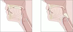
Background imageGullet Collection: Mechanics of swallowing, diagram

Mechanics of swallowing, diagram
Mechanics of swallowing. Cutaway diagrams showing the process of swallowing. At left the mouthful of food (the bolus, green) is being pushed to the back of the mouth by the tongue

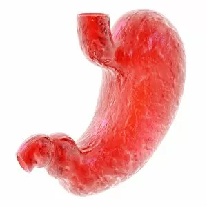
Background imageGullet Collection: Stomach, computer artwork

Stomach, computer artwork
Stomach. Computer artwork of the stomach, the first major organ of the digestive system. Food enters the stomach from the oesophagus (upper centre)

Background imageGullet Collection: Oesophagus, X-ray

Oesophagus, X-ray
Oesophagus, coloured X-ray. The oesophagus (orange) is the tube that carries food and drink from the mouth to the stomach. It has been visualised here using a barium swallow

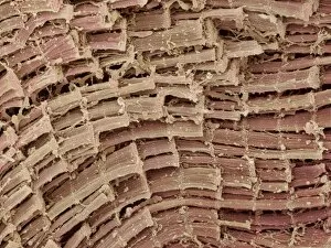
Background imageGullet Collection: Oesophagus, SEM

Oesophagus, SEM
Oesophagus. Coloured scanning electron micrograph (SEM) of a freeze-fracture through the muscularis externa of the oesophagus. This forms part of the muscularis mucosa found throughout the gut

Background imageGullet Collection: Oesophagus epithelium, SEM

Oesophagus epithelium, SEM
Oesophagus epithelium. Coloured scanning electron micrograph (SEM) of the lining of the human oesophagus or gullet (yellow) and two unknown bacteria (pink & purple)

Background imageGullet Collection: Oesophagus lining, SEM

Oesophagus lining, SEM
Oesophagus lining. Coloured scanning electron micrograph (SEM) of the lining (epithelium) of the human oesophagus (gullet, blue) and two bacteria (red)

Background imageGullet Collection: Heartburn

Heartburn. Computer artwork of stomach acid in the oesophagus, the tube that leads from the mouth to the stomach, the cause of heartburn

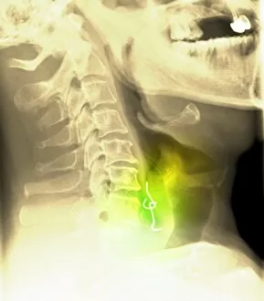
Background imageGullet Collection: Swallowed denture, X-ray

Swallowed denture, X-ray

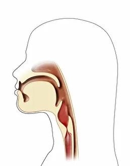
Background imageGullet Collection: Mouth and throat anatomy, artwork

Mouth and throat anatomy, artwork

Background imageGullet Collection: Human internal organs, diagram

Human internal organs, diagram
Human internal organs. Diagram showing the human internal organs in the head and torso. In the head, the organs and cavities shown are: the brain (encephalon), the cerebellum (part of the brain)

Background imageGullet Collection: Human internal organs, artwork

Human internal organs, artwork
Human internal organs. Artwork showing the human internal organs in the head and torso. In the head, the organs and cavities shown are: the brain (encephalon), the cerebellum (part of the brain)

Background imageGullet Collection: Upper digestive system, artwork

Upper digestive system, artwork
Upper digestive system, computer artwork. At top is the mouth, from which food passes into the oesophagus (gullet), a tube that leads down into the stomach

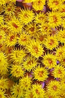
Background imageGullet Collection: Coral

Coral. Close-up of the coralTubastrea sp. Corals consist of colonies of individual animals called polyps. The polyps are simple creatures, with a ring of tentacles leading to a gullet

Background imageGullet Collection: Gulet anchored at Kalkan, a popular tourist resort, Antalya Province, Anatolia

Gulet anchored at Kalkan, a popular tourist resort, Antalya Province, Anatolia, Turkey, Asia Minor, Eurasia

Background imageGullet Collection: Gullet anchored at Kalkan, a popular tourist resort, Antalya Province, Anatolia

Gullet anchored at Kalkan, a popular tourist resort, Antalya Province, Anatolia, Turkey, Asia Minor, Eurasia


For sale as Licensed Images

Choose your image, Select your licence and Download the media

"The Gullet: A Journey Through Time and Culture" Step into the historical charm of Stafford Street, Birmingham, where "The Gullet" stands as a testament to the city's rich heritage. As you enter this vibrant street, prepare to embark on a captivating adventure that spans across various eras and continents. Immerse yourself in artistry with the Pyrenean Woman force-feeding a goose painting. This masterpiece transports you to an era when creativity knew no bounds. The intricate details and vivid colors evoke emotions that transcend time. Continuing our journey through history, we encounter Midas, Transmuting all into [Gold] Paper. This hand-colored engraving from 1797 captivates with its depiction of Midas' legendary power. It serves as a reminder of humanity's fascination with wealth and its consequences. From ancient legends to modern-day wonders, we set sail towards Kalkan in Anatolia aboard a magnificent Gulet anchored at this popular tourist resort. Feel the gentle breeze caress your face as you admire the breathtaking beauty of Antalya Province. Delve deeper into knowledge by exploring anatomical wonders such as the male respiratory system and internal organs alongside their female counterparts. These illustrations offer fascinating insights into our complex biology while showcasing the intricacies of human life. Jacques Fabien Gautier Dagoty's Two Views of the Head takes us back to 1746 France, where artistic mastery met scientific curiosity. Marvel at how these detailed renderings capture both external features and hidden depths within each subject's visage. As our journey nears its end, Petra beckons with its enigmatic allure captured in black-and-white photographs featuring gullets and bridges alike. Let your imagination wander amidst these ancient ruins; feel their stories whisper through time itself. Reflect upon societal changes depicted in Hannah Humphrey's engravings like "Birmingham Improvements under Artisans and Labourers Dwellings Improvement Act, 1875.