
Haematological Collection (#4)

"Exploring the Intricate World Marvels

Background imageHaematological Collection: Normal and leukaemic blood, artwork C016 / 6261

Normal and leukaemic blood, artwork C016 / 6261
Normal and leukaemic blood. Computer artwork comparing the appearance of normal blood (left) and that affected by leukaemia (right)

Background imageHaematological Collection: Red blood cells, SEM C015 / 8789

Red blood cells, SEM C015 / 8789
Red blood cells. Coloured scanning electron micrograph (SEM) of human red blood cells (erythrocytes). Red blood cells are biconcave, giving them a large surface area for gas exchange

Background imageHaematological Collection: Red blood cells, SEM C015 / 8792

Red blood cells, SEM C015 / 8792
Red blood cells. Coloured scanning electron micrograph (SEM) of human red blood cells (erythrocytes). Red blood cells are biconcave, giving them a large surface area for gas exchange

Background imageHaematological Collection: Red blood cells, SEM C015 / 8794

Red blood cells, SEM C015 / 8794
Red blood cells. Coloured scanning electron micrograph (SEM) of human red blood cells (erythrocytes). Red blood cells are biconcave, giving them a large surface area for gas exchange

Background imageHaematological Collection: Red blood cells, SEM C015 / 8796

Red blood cells, SEM C015 / 8796
Red blood cells. Coloured scanning electron micrograph (SEM) of human red blood cells (erythrocytes). Red blood cells are biconcave, giving them a large surface area for gas exchange

Background imageHaematological Collection: Red blood cells, SEM C015 / 8790

Red blood cells, SEM C015 / 8790
Red blood cells. Coloured scanning electron micrograph (SEM) of human red blood cells (erythrocytes). Red blood cells are biconcave, giving them a large surface area for gas exchange

Background imageHaematological Collection: Thrombin complexed with fibrinogen C015 / 7148

Thrombin complexed with fibrinogen C015 / 7148
Thrombin complexed with fibrinogen, molecular model. The thrombin molecules (left and right, purple and green) are bound to the central part of the fibrinogen molecule (centre, multiple colours)

Background imageHaematological Collection: Haemoglobin, molecular model C015 / 8938

Haemoglobin, molecular model C015 / 8938
Haemoglobin, molecular model. This molecule transports oxygen around the body in red blood cells. It consists of four globin proteins (amino acid chains; orange, green, blue and purple)

Background imageHaematological Collection: Reticulosarcoma, light micrograph C015 / 7133

Reticulosarcoma, light micrograph C015 / 7133
Reticulosarcoma. Light micrograph of reticulocyte blood cells (red, one at upper left) from a lymphatic ganglion in a case of reticulosarcoma

Background imageHaematological Collection: Red blood cells, SEM C015 / 8793

Red blood cells, SEM C015 / 8793
Red blood cells. Coloured scanning electron micrograph (SEM) of human red blood cells (erythrocytes). Red blood cells are biconcave, giving them a large surface area for gas exchange

Background imageHaematological Collection: Haemoglobin, molecular model C015 / 9391

Haemoglobin, molecular model C015 / 9391
Haemoglobin, molecular model. This is deoxyhaemoglobin, the molecule in its non-oxygen bound state. Haemoglobin transports oxygen around the body in red blood cells

Background imageHaematological Collection: Red blood cells, SEM C015 / 8795

Red blood cells, SEM C015 / 8795
Red blood cells. Coloured scanning electron micrograph (SEM) of human red blood cells (erythrocytes). Red blood cells are biconcave, giving them a large surface area for gas exchange

Background imageHaematological Collection: Haemoglobin, molecular model C015 / 8939

Haemoglobin, molecular model C015 / 8939
Haemoglobin, molecular model. This molecule transports oxygen around the body in red blood cells. It consists of four globin proteins (amino acid chains; orange, green, blue and purple)

Background imageHaematological Collection: Red blood cells, SEM C015 / 8787

Red blood cells, SEM C015 / 8787
Red blood cells. Coloured scanning electron micrograph (SEM) of human red blood cells (erythrocytes). Red blood cells are biconcave, giving them a large surface area for gas exchange

Background imageHaematological Collection: Red blood cells, SEM C015 / 8791

Red blood cells, SEM C015 / 8791
Red blood cells. Coloured scanning electron micrograph (SEM) of human red blood cells (erythrocytes). Red blood cells are biconcave, giving them a large surface area for gas exchange

Background imageHaematological Collection: Red blood cells, SEM C015 / 8788

Red blood cells, SEM C015 / 8788
Red blood cells. Coloured scanning electron micrograph (SEM) of human red blood cells (erythrocytes). Red blood cells are biconcave, giving them a large surface area for gas exchange

Background imageHaematological Collection: Blood cells, light micrograph C015 / 7130

Blood cells, light micrograph C015 / 7130
Blood cells. Light micrograph of red blood cells (erythrocytes, red) and white blood cells (leucocytes, nuclei stained pink)

Background imageHaematological Collection: Thrombin complexed with fibrinogen C015 / 7149

Thrombin complexed with fibrinogen C015 / 7149
Thrombin complexed with fibrinogen, molecular model. The thrombin molecules (left and right, brown and pink) are bound to the central part of the fibrinogen molecule (centre, multiple colours)

Background imageHaematological Collection: Haemoglobin, molecular model C015 / 9392

Haemoglobin, molecular model C015 / 9392
Haemoglobin, molecular model. This is deoxyhaemoglobin, the molecule in its non-oxygen bound state. Haemoglobin transports oxygen around the body in red blood cells

Background imageHaematological Collection: Malaria infection cycle, artwork

Malaria infection cycle, artwork. Life cycle of the Plasmodium parasite that causes malaria, which can cause coma and death. At upper left, a female Anopheles mosquito bites and infects a person

Background imageHaematological Collection: Blood cells, illustration C018 / 0802

Blood cells, illustration C018 / 0802
Blood cells. All cellular blood components originate from the same cell, the haematopoietic stem cell. The stem cell differentiates into two types of progenitor cells

Background imageHaematological Collection: Blood clot, SEM C014 / 0381

Blood clot, SEM C014 / 0381
Blood clot. Coloured scanning electron micrograph (SEM) of a blood clot (thrombus) in an arteriole (small blood vessel) of a salivary gland

Background imageHaematological Collection: Monocyte white blood cell, TEM

Monocyte white blood cell, TEM
Monocyte white blood cell. Transmission electron micrograph (TEM) of a section through a monocyte white blood cell, showing the cell nucleus (blue)

Background imageHaematological Collection: Blood clot, SEM C014 / 0380

Blood clot, SEM C014 / 0380
Blood clot. Coloured scanning electron micrograph (SEM) of a blood clot (thrombus) in an arteriole (small blood vessel) of a salivary gland

Background imageHaematological Collection: Red blood cells, artwork C016 / 8542

Red blood cells, artwork C016 / 8542
Red blood cells in a blood vessel, computer artwork. Red blood cells are biconcave, giving them a large surface area for gas exchange, and highly elastic

Background imageHaematological Collection: Red blood cells, artwork C016 / 8547

Red blood cells, artwork C016 / 8547
Red blood cells in a blood vessel, computer artwork. Red blood cells are biconcave, giving them a large surface area for gas exchange, and highly elastic

Background imageHaematological Collection: Red blood cells, artwork C016 / 8543

Red blood cells, artwork C016 / 8543
Red blood cells in a blood vessel, computer artwork. Red blood cells are biconcave, giving them a large surface area for gas exchange, and highly elastic

Background imageHaematological Collection: Red blood cells, artwork C016 / 8548

Red blood cells, artwork C016 / 8548
Red blood cells in a blood vessel, computer artwork. Red blood cells are biconcave, giving them a large surface area for gas exchange, and highly elastic

Background imageHaematological Collection: Red blood cells, artwork C016 / 8546

Red blood cells, artwork C016 / 8546
Red blood cells in a blood vessel, computer artwork. Red blood cells are biconcave, giving them a large surface area for gas exchange, and highly elastic

Background imageHaematological Collection: Red blood cells, artwork C016 / 8544

Red blood cells, artwork C016 / 8544
Red blood cells in a blood vessel, computer artwork. Red blood cells are biconcave, giving them a large surface area for gas exchange, and highly elastic

Background imageHaematological Collection: Kidney blood vessels, confocal micrograph C014 / 4610

Kidney blood vessels, confocal micrograph C014 / 4610
Kidney blood vessels. Laser scanning confocal micrograph (LSCM) of a section through a human kidney, showing a blood vessel (vertical, centre)

Background imageHaematological Collection: Blood cells, SEM C016 / 8026

Blood cells, SEM C016 / 8026
Blood cells. Coloured scanning electron micrograph (SEM) of a lymphocyte white blood cell (centre) and red blood cells (erythrocytes, circular)

Background imageHaematological Collection: Red blood cells, SEM C016 / 8027

Red blood cells, SEM C016 / 8027
Red blood cells. Coloured scanning electron micrograph (SEM) of red blood cells (erythrocytes). The main function of red blood cells is to distribute oxygen to body tissues

Background imageHaematological Collection: Blood vessels, light micrograph C014 / 4562

Blood vessels, light micrograph C014 / 4562
Blood vessels. Light micrograph of a section through healthy human tissue showing an artery (large, red) and vein (small, red), surrounded by connective tissue (yellow)

Background imageHaematological Collection: Red blood cells, artwork C013 / 5932

Red blood cells, artwork C013 / 5932
Red blood cells (erythrocytes), computer artwork

Background imageHaematological Collection: Haemoglobin S, molecular model

Haemoglobin S, molecular model
Haemoglobin S. Molecular model of the mutant form of haemoglobin (haemoglobin S) that causes sickle cell anaemia. This is deoxyhaemoglobin S, the molecule in its non-oxygen bound state

Background imageHaematological Collection: Blood sample cryostorage C015 / 6530

Blood sample cryostorage C015 / 6530
Blood sample cryostorage. Close-up of a freezer used to store samples of human blood for use in research in a molecular epidemiology lab. This freezer holds the samples at -20 degrees Celsius

Background imageHaematological Collection: Blood sample cryostorage C015 / 6531

Blood sample cryostorage C015 / 6531
Blood sample cryostorage. Close-up of a freezer used to store samples of human blood for use in research in a molecular epidemiology lab. This freezer holds the samples at -20 degrees Celsius

Background imageHaematological Collection: Blood sample cryostorage C015 / 6529

Blood sample cryostorage C015 / 6529
Blood sample cryostorage. Close-up of a freezer used to store samples of human blood for use in research in a molecular epidemiology lab. This freezer holds the samples at -20 degrees Celsius

Background imageHaematological Collection: Blood sampling C015 / 3466

Blood sampling C015 / 3466
Blood sampling. Blood sample being taken from a patients arm. The sample of blood will be labelled and sent for testing. Blood is analysed for levels of components such as cholesterol and glucose

Background imageHaematological Collection: Blood sampling C015 / 3465

Blood sampling C015 / 3465
Blood sampling. Blood sample being taken from a patients arm. The sample of blood will be labelled and sent for testing. Blood is analysed for levels of components such as cholesterol and glucose

Background imageHaematological Collection: Healthy artery, artwork C015 / 6444

Healthy artery, artwork C015 / 6444
Healthy artery. Computer artwork of the inside of an healthy artery, showing red blood cells (circular) and the epithelial cells (square) lining the vessels wall

Background imageHaematological Collection: Sickle cell anaemia, light micrograph C015 / 6395

Sickle cell anaemia, light micrograph C015 / 6395
Sickle cell anaemia. Light micrograph of a blood smear showing normally rounded red blood cells that have become curved, elongated sickle-shaped cells

Background imageHaematological Collection: Uterine bleeding treatment, DSA scan

Uterine bleeding treatment, DSA scan
Uterine bleeding treatment. Coloured digital subtraction angiogram (DSA) of the pelvic region of a 45-year-old woman with menometrorrhagia, showing embolisation of the uterine arteries

Background imageHaematological Collection: Red and white blood cells, artwork C016 / 4629

Red and white blood cells, artwork C016 / 4629
Red and white blood cells, computer artwork. Red blood cells are biconcave, giving them a large surface area for gas exchange, and highly elastic

Background imageHaematological Collection: Red and white blood cells, artwork C016 / 4630

Red and white blood cells, artwork C016 / 4630
Red and white blood cells, computer artwork. Red blood cells are biconcave, giving them a large surface area for gas exchange, and highly elastic

Background imageHaematological Collection: Red blood cells, artwork C016 / 4627

Red blood cells, artwork C016 / 4627
Red blood cells, computer artwork. Red blood cells are biconcave, giving them a large surface area for gas exchange, and highly elastic, enabling them to pass through narrow capillary vessels

Background imageHaematological Collection: Red blood cells, artwork C016 / 4626

Red blood cells, artwork C016 / 4626
Red blood cells, computer artwork. Red blood cells are biconcave, giving them a large surface area for gas exchange, and highly elastic, enabling them to pass through narrow capillary vessels


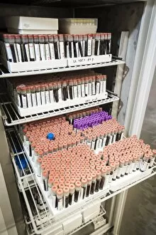
For sale as Licensed Images

Choose your image, Select your licence and Download the media

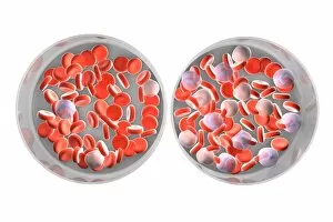
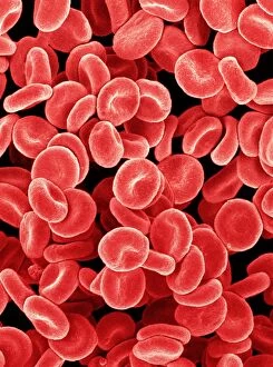
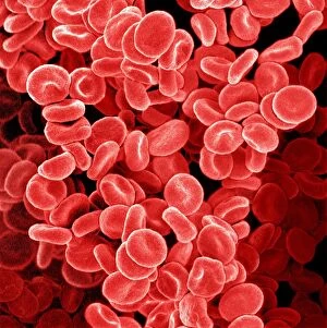
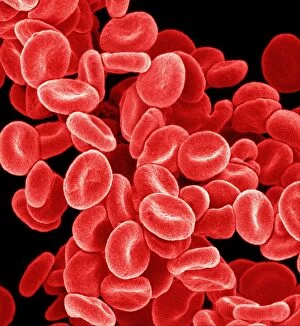
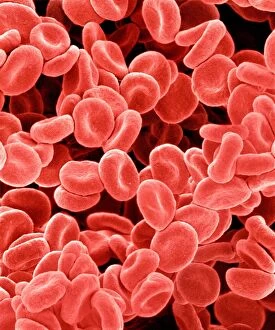
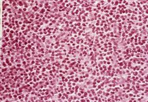
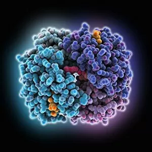
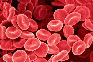
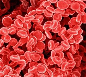
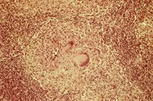
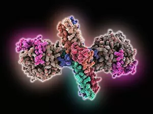
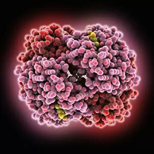
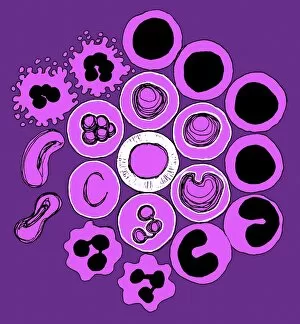
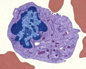
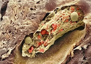
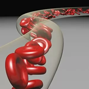
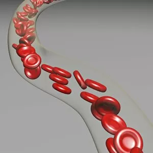
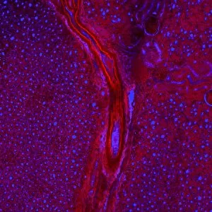
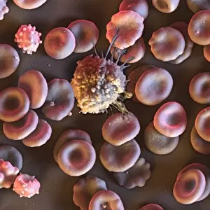
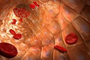
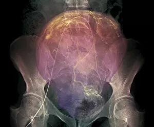
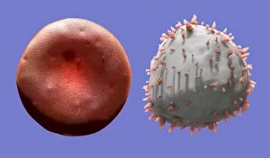
"Exploring the Intricate World Marvels: From Blood Coagulation to Leukaemia" Delve into the mesmerizing realm of haematology with a captivating artwork showcasing the intricate blood coagulation cascade (Artwork C016 / 9873). Witness the enigmatic Dohle bodies within a blood cell through a powerful micrograph, unraveling their significance in haematological diagnostics. Behold the stunning beauty of red blood cells captured by a scanning electron microscope (SEM), revealing their unique structure and function. Uncover the haunting reality of Acute Promyelocytic Leukaemia through an evocative micrograph, shedding light on this devastating blood disorder. Immerse yourself in an artistic portrayal of lymphocyte white blood cells, celebrating their vital role in our immune system's defense against pathogens (Artwork). Get up close and personal with a striking SEM image capturing the intricate details of a blood clot formation, highlighting its crucial role in wound healing and preventing excessive bleeding. Explore the fascinating world of haematopoietic stem cells through a remarkable SEM image (C013 / 5009), marveling at their ability to give rise to all types of mature blood cells. Journey into the microscopic realm as you witness myeloblasts and promyelocytes under light microscopy, providing insights into early stages of white blood cell development. Peer into an extraordinary TEM image depicting macrophages interacting with lymphocytes, offering glimpses into how these immune cells collaborate for optimal defense against infections. Experience awe-inspiring SEM images capturing white blood cells alongside platelets (C016 / 3099 & C016 / 3098), illustrating their collective effort in maintaining our body's delicate balance between immunity and clotting mechanisms.