
Hair Follicle Collection
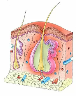
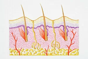
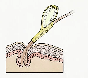
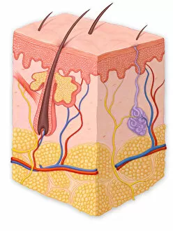
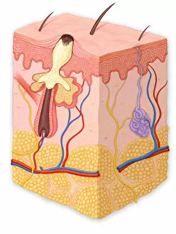
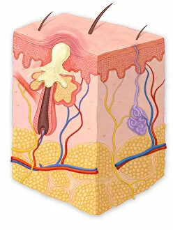
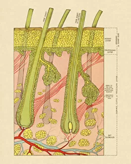
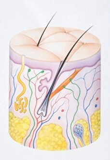
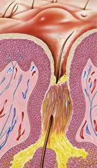
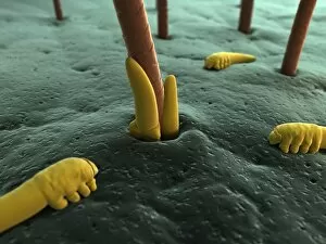
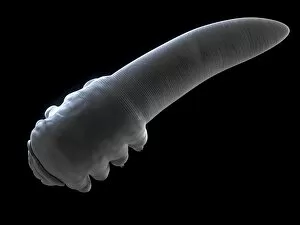
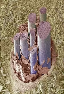
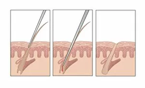
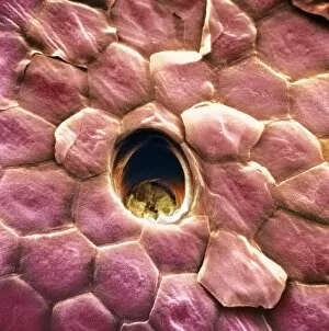
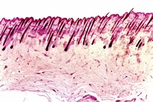
The hair follicle: a microscopic world within our skin


For sale as Licensed Images
Choose your image, Select your licence and Download the media
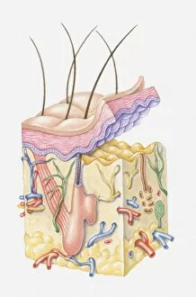
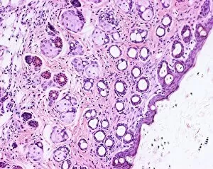
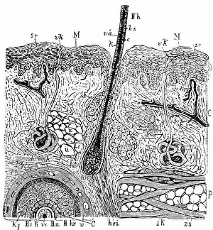
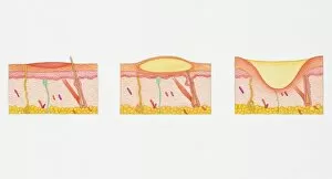
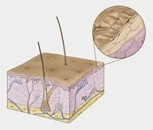
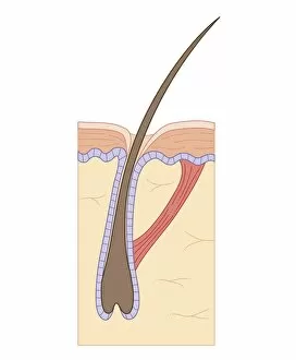
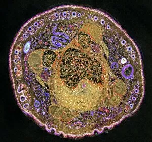
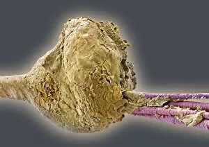
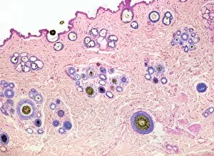
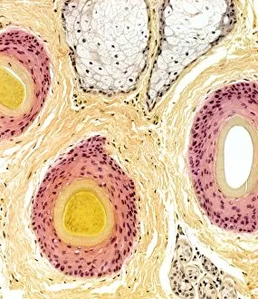
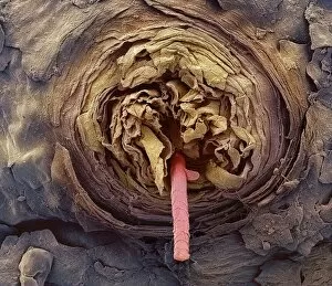
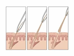
The hair follicle: a microscopic world within our skin. 🌟 Step into the fascinating realm of human hair follicles and skin, where intricate processes unfold to create our luscious locks. In this captivating journey, we explore various aspects that make up this hidden wonderland. Firstly, let's delve into the cross-section of human skin with heat trapped by erect hairs. This illustration showcases how our body reacts to cold temperatures, causing tiny muscles around the hair follicles to contract and raise the hairs on our skin – an instinctive response known as "goosebumps. " Moving on, we encounter the eyelash follicle in stunning detail through scanning electron microscopy (SEM). Witnessing its delicate structure reminds us of nature's precision in crafting even the tiniest features. As we venture deeper into normal cross-sections of the skin in layers, we uncover its complex composition. From epidermis to dermis and intertwined within them – like roots anchoring a tree – are countless hair follicles that nurture each strand from birth until it graces our heads. But not all is smooth sailing; sometimes blemishes arise. A blackhead emerges when excess oil clogs a pore while still exposed to air. On another path lies a papule—a small red bump signaling inflammation beneath the surface—reminding us of our body's constant battle against impurities. Further along this expedition through skin cross sections awaits a pustule—an inflamed lesion filled with pus—a stark reminder that even within beauty lies imperfection. And nearby stands a whitehead—a closed comedo formed when sebum blocks a pore entirely—showcasing yet another facet of this intricate ecosystem. Finally, behold an enchanting diagram illustrating two layers: epidermis and dermis alongside their faithful companion—the hair follicle. Together they form an inseparable bond responsible for maintaining healthy strands throughout life's changes.