Helices Collection (#2)

"Unraveling the Mysteries of Helices: Exploring the Intricate World of Molecular Structures" DNA Transcription Unveiled

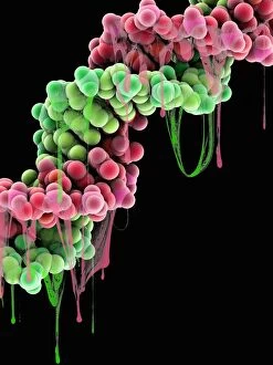
Background imageHelices Collection: Damaged DNA, conceptual artwork C013 / 9999

Damaged DNA, conceptual artwork C013 / 9999
Damaged DNA, conceptual computer artwork

Background imageHelices Collection: Human growth hormone molecule C013 / 9006

Human growth hormone molecule C013 / 9006
Human growth hormone. Molecular model of human growth hormone (hGH, yellow) bound to the extracellular domain of the human growth hormone binding protein (hGHBP)

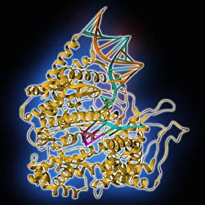
Background imageHelices Collection: RNA polymerase molecule C013 / 9005

RNA polymerase molecule C013 / 9005
RNA polymerase. Molecular model of RNA polymerase (yellow) transcribing a strand of mRNA (messenger ribonucleic acid, pink) from a DNA (deoxyribonucleic acid) template (orange and turquoise)

Background imageHelices Collection: HIV reverse transcription enzyme C013 / 8998

HIV reverse transcription enzyme C013 / 8998
HIV reverse transcription enzyme. Molecular model of the reverse transcriptase enzyme (orange and blue) found in HIV (the human immunodeficiency virus)

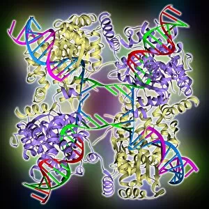
Background imageHelices Collection: DNA Holliday junction complex C013 / 8888

DNA Holliday junction complex C013 / 8888
DNA Holliday junction complex. Molecular model of the enzyme FLP recombinase in complex with a Holliday junction between homologous strands of DNA (deoxyribonucleic acid)

Background imageHelices Collection: Erythropoietin molecule C013 / 8891

Erythropoietin molecule C013 / 8891
Erythropoietin molecule (EPO), molecular model. EPO is a glycoprotein that is produced by the kidneys in response to low blood oxygen levels

Background imageHelices Collection: Birch pollen allergen molecule C013 / 8889

Birch pollen allergen molecule C013 / 8889
Birch pollen allergen molecule. Computer model showing the secondary structure of a Bet v 1L molecule. This molecule is responsible for allergic reactions to pollen from birch (Betula sp.) trees

Background imageHelices Collection: Oxoguanine glycosylase complex C013 / 8886

Oxoguanine glycosylase complex C013 / 8886
Oxoguanine glycosylase complex. Computer model showing a molecule of human aG DNA repair glycosylase (right) bound to an DNA molecule (left)

Background imageHelices Collection: Oxoguanine glycosylase complex C013 / 8884

Oxoguanine glycosylase complex C013 / 8884
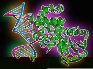
Oxoguanine glycosylase complex. Computer model showing an 8-Oxoguanine glycosylase (OGG1) molecule (green) bound to a section of DNA (deoxyribonucleic acid, pink and blue)

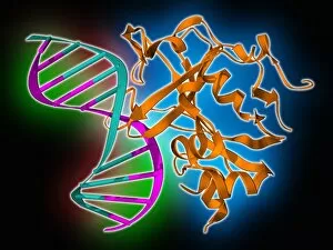
Background imageHelices Collection: TATA box-binding protein complex C013 / 8881

TATA box-binding protein complex C013 / 8881
TATA box-binding protein complex. Molecular model showing a TATA box-binding protein (TBP) (purple) complexed with a strand of DNA (deoxyribonucleic acid, blue)

Background imageHelices Collection: Potassium channel molecule C013 / 8878

Potassium channel molecule C013 / 8878
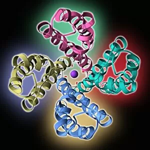
Potassium channel molecule. Computer model showing the secondary structure of a KcsA potassium ion (K+) channel molecule from Streptomyces lividans bacteria

Background imageHelices Collection: Bovine adrenodoxin molecules C013 / 8876

Bovine adrenodoxin molecules C013 / 8876
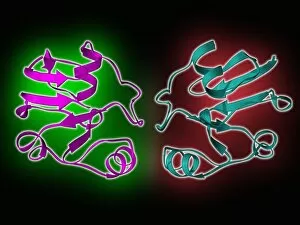
Bovine adrenodoxin molecules. Computer models showing the secondary structure of two molecules of adrenodoxin (adrenal ferredoxin) from a cow

Background imageHelices Collection: Cytochrome b5 molecules C013 / 8874

Cytochrome b5 molecules C013 / 8874
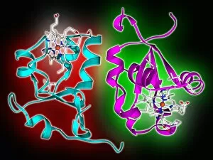
Cytochrome b5 molecules. Computer artwork showing the secondary structure of two molecules of cytochrome B5. This protein plays a crucial role in metabolism in animals (including humans), fungi

Background imageHelices Collection: Pit-1 transcription factor bound to DNA C013 / 8872

Pit-1 transcription factor bound to DNA C013 / 8872
Pit-1 transcription factor bound to DNA. Molecular model showing pituitary-specific positive transcription factor 1 (Pit-1) (purple and yellow) bound to a strand of DNA (deoxyribonucleic acid)

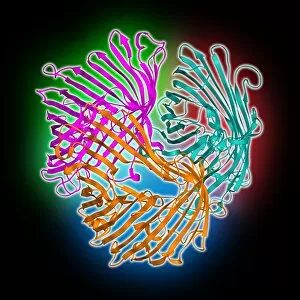
Background imageHelices Collection: Sucrose-specific porin molecule C013 / 8870

Sucrose-specific porin molecule C013 / 8870
Sucrose-specific porin molecule. Moleuclar model showing the secondary and quaternary structure of a molecule of sucrose-specific porin

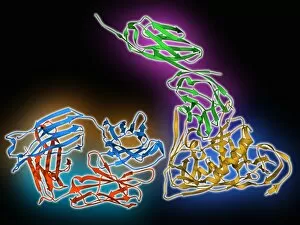
Background imageHelices Collection: HIV antibody therapy, molecular model C013 / 7908

HIV antibody therapy, molecular model C013 / 7908
HIV antibody therapy. Molecular model of the interaction of the HIV surface protein gp120 (yellow, bottom right) as it interacts with a human white blood cell surface protein (CD4, green)

Background imageHelices Collection: Glycated haemoglobin molecule C013 / 7781

Glycated haemoglobin molecule C013 / 7781
Glycated haemoglobin molecule. Computer model of a glycated haemoglobin molecule. The alpha and beta subunits of the haemoglobin are blue and pink, and the iron-containing haem groups are grey

Background imageHelices Collection: Glycated haemoglobin molecule C013 / 7779

Glycated haemoglobin molecule C013 / 7779
Glycated haemoglobin molecule. Computer model showing a glucose molecule (centre) bound to a molecule of haemoglobin. The alpha and beta subunits of the haemoglobin are blue and pink

Background imageHelices Collection: Projet d appareil, pour evaluer approximativement la force d

Projet d appareil, pour evaluer approximativement la force des helices, roues ailes applicables e la direction des a erostat

Background imageHelices Collection: Genetic sexuality

Genetic sexuality. Conceptual computer artwork of male and female symbols in front of DNA helices (deoxyribonucleic acid), representing the genetic component of sexual identity

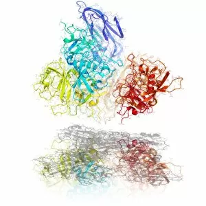
Background imageHelices Collection: Bluetongue virus core protein

Bluetongue virus core proteins, molecular model. Bluetongue virus (BTV) is an Orbivirus that causes a potentially fatal disease in sheep, cattle and other ruminants. It does not affect humans

Background imageHelices Collection: Norwalk virus capsid protein

Norwalk virus capsid protein. Molecular model showing the secondary structure of the capsid protein from a norwalk virus particle

Background imageHelices Collection: GM plants, conceptual artwork

GM plants, conceptual artwork
Genetically modified plants, conceptual artwork. Plant stems and leaves forming structures that echo DNA (deoxyribonucleic acid) double helices. This could represent genetically modified (GM) plants

Background imageHelices Collection: DNA with money

DNA with money. Computer artwork of a DNA double helix superimposed over a British 20 pound note. This image could represent the commercial implications of DNA research

Background imageHelices Collection: Destruction of DNA helix

Destruction of DNA helix. The computer artwork may represent the breakdown of DNA during an explosion or the destruction of DNA in hot, molten lava

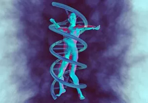
Background imageHelices Collection: Man and DNA

Man and DNA. Computer artwork of a DNA molecule wrapped around a male figure. A molecule of DNA (deoxyribonucleic acid) consists of two strands of sugar phosphates forming a double helix (spiral)

Background imageHelices Collection: DNA recombination, molecular model

DNA recombination, molecular model
DNA recombination. Molecular model of a Holliday Junction, a moving point of contact between two DNA (deoxyribonucleic acid)

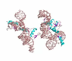
Background imageHelices Collection: Zinc finger-RNA complex

Zinc finger-RNA complex. Computer models of zinc finger protein molecules complexed with RNA (ribonucleic acid) molecules

Background imageHelices Collection: DNA in bubble

DNA in bubble. Conceptual computer artwork of a DNA double helix within a bubble in water. This image could represent the fragility of the DNA molecule or the need for protection of an individuals

Background imageHelices Collection: DNA molecules, conceptual artwork

DNA molecules, conceptual artwork
DNA molecules, conceptual computer artwork

Background imageHelices Collection: DNA in space

DNA in space. Computer artwork of a DNA double helix in space above Earth. This may represent the possible uniqueness of Earth as a cradle of life in the universe


For sale as Licensed Images

Choose your image, Select your licence and Download the media

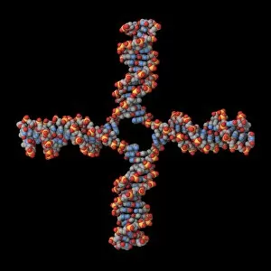
"Unraveling the Mysteries of Helices: Exploring the Intricate World of Molecular Structures" DNA Transcription Unveiled: Witness the intricate dance as DNA unravels and transcribes its genetic code, paving the way for life's blueprint. A Symphony in Proteins: Secondary structures adorned with helical motifs create a mesmerizing molecular model, showcasing nature's artistic prowess. Nucleosome Molecule - Nature's Genetic Sculptor: Behold the elegant architecture of nucleosomes, where DNA elegantly wraps around histone proteins forming a helical masterpiece. The Dance of Life: An artwork depicting a vibrant DNA molecule reveals its double-helix structure, symbolizing our very essence and genetic heritage. Bacterial Ribosome - The Protein Factory: Dive into the intricacies of bacterial ribosomes as they diligently translate genetic information into functional proteins through their complex helical framework. HIV Reverse Transcription Enzyme - Decoding Viral Secrets: Explore how this remarkable enzyme utilizes helical structures to reverse-transcribe viral RNA into infectious DNA, perpetuating HIV's stealthy invasion. Hepatitis C Virus Enzyme - Unmasking an Invisible Threat: Peer inside the molecular model of this cunning enzyme that hijacks human cells by employing intricate helices to replicate itself relentlessly. Interferon Molecule - Our Body's Defender: Discover how these mighty molecules employ their unique helical shape to activate our immune system against invading pathogens and protect us from harm. Z-DNA Tetramer Molecule C015/6557 – Unlocking New Dimensions in Genetics: Delve into the fascinating world of Z-DNA tetramers as they challenge conventional double-helical structures, offering new insights into gene regulation and potential therapeutic avenues. Cholera Toxin – A Deadly Ballet on a Molecular Stage.