
Hip Bone Collection (#2)

The hip bone, a vital component of the pelvis, plays a crucial role in our daily activities

Background imageHip Bone Collection: Leg anatomy, artwork C013 / 4494

Leg anatomy, artwork C013 / 4494
Leg anatomy. Computer artwork of an oblique anterior view looking down a human leg, showing muscles and tendons (brown and white), bones and ligaments (white), arteries and veins (red and blue)

Background imageHip Bone Collection: Leg ligaments, artwork

Leg ligaments, artwork
Leg anatomy. Computer artwork of an anterior view of a human leg, showing the muscles and tendons (brown and white), bones and ligaments (white), arteries and veins (red and blue), nerves (yellow)

Background imageHip Bone Collection: Hip ligaments, artwork C013 / 4434

Hip ligaments, artwork C013 / 4434
Hip ligaments. Computer artwork of a posterior oblique view of the hip bones and ligaments (white). At upper right the lumbar spine ends in the sacrum and coccyx (tailbone)

Background imageHip Bone Collection: Hip ligaments, artwork C013 / 4433

Hip ligaments, artwork C013 / 4433
Hip ligaments. Computer artwork of a posterior view of the hip bones and ligaments (white). At centre the lumbar spine ends in the sacrum and coccyx (tailbone)

Background imageHip Bone Collection: Hip ligaments, artwork C013 / 4432

Hip ligaments, artwork C013 / 4432
Hip ligaments. Computer artwork of an anterior view of the hip bones and ligaments (white). At centre the lumbar spine ends in the sacrum and coccyx (tailbone)

Background imageHip Bone Collection: Hip anatomy, artwork C013 / 4430

Hip anatomy, artwork C013 / 4430
Hip anatomy. Computer artwork of the head of the left femur (centre right) articulating with the left-hand side of the pelvis to form the hip joint

Background imageHip Bone Collection: Leg muscles, artwork C013 / 4429

Leg muscles, artwork C013 / 4429
Leg muscles. Computer artwork of the bones and outer muscles of the left leg, seen from the front (anterior view) at an oblique angle looking downwards

Background imageHip Bone Collection: Thigh hamstring muscles, artwork C013 / 4428

Thigh hamstring muscles, artwork C013 / 4428
Thigh hamstring muscles. Computer artwork of a posterior view of two of the three hamstring muscles (red) of the thighs. The muscles are the semimembranosus (inner) and semitendinosus (outer)

Background imageHip Bone Collection: Iliopsoas muscles, artwork C013 / 4425

Iliopsoas muscles, artwork C013 / 4425
Iliopsoas muscles. Computer artwork of an anterior (frontal) view of the hips, showing the inner hip muscles (red), the ilioposas

Background imageHip Bone Collection: Buttock muscles, artwork C013 / 4423

Buttock muscles, artwork C013 / 4423
Buttock muscles. Computer artwork of a posterior oblique view of the hips, showing some of the muscles (red) of the buttocks

Background imageHip Bone Collection: Buttock muscles, artwork C013 / 4424

Buttock muscles, artwork C013 / 4424
Buttock muscles. Computer artwork of a posterior view of the hips showing some of the muscles (red) of the buttocks. These are shown attached to the femurs (thigh bones) and the pelvis bones

Background imageHip Bone Collection: Buttock muscles, artwork C013 / 4422

Buttock muscles, artwork C013 / 4422
Buttock muscles. Computer artwork of a posterior oblique view of the hips showing some of the muscles (red) of the buttocks. These are shown attached to the femurs (thigh bones) and the pelvis bones

Background imageHip Bone Collection: Buttock muscles, artwork C013 / 4420

Buttock muscles, artwork C013 / 4420
Buttock muscles. Computer artwork of a posterior view of the hips showing some of the muscles (red) of the buttocks. These are shown attached to the femurs (thigh bones) and the pelvis bones

Background imageHip Bone Collection: Buttock muscles, artwork C013 / 4421

Buttock muscles, artwork C013 / 4421
Buttock muscles. Computer artwork of a side view (front of body at left) of the hips showing some of the muscles (red) of the buttocks

Background imageHip Bone Collection: Buttock muscles, artwork C013 / 4419

Buttock muscles, artwork C013 / 4419
Buttock muscles. Computer artwork of a posterior oblique view of the hips showing some of the muscles (red) of the buttocks. These are shown attached to the femurs (thigh bones) and the pelvis bones

Background imageHip Bone Collection: Buttock muscles, artwork C013 / 4415

Buttock muscles, artwork C013 / 4415
Buttock muscles. Computer artwork of a posterior oblique view of the hips showing some of the muscles (red) of the buttocks. These are shown attached to the femurs (thigh bones) and the pelvis bones

Background imageHip Bone Collection: Buttock muscles, artwork C013 / 4416

Buttock muscles, artwork C013 / 4416
Buttock muscles. Computer artwork of a posterior oblique view of the hips showing some of the muscles (red) of the buttocks. These are shown attached to the femurs (thigh bones) and the pelvis bones

Background imageHip Bone Collection: Spine, computer artwork

Spine, computer artwork
Spine. Computer artwork showing the bones of the spine, shoulder and pelvis on the human body. The spine, or backbone, supports the back and protects the spinal cord

Background imageHip Bone Collection: Mammoth bone musical instruments

Mammoth bone musical instruments
Prehistoric carved mammoth bone musical instruments. Dating from around 20, 000 years ago, during the Stone Age, these carvings are believed to have been used by early Man to produce sound


For sale as Licensed Images

Choose your image, Select your licence and Download the media

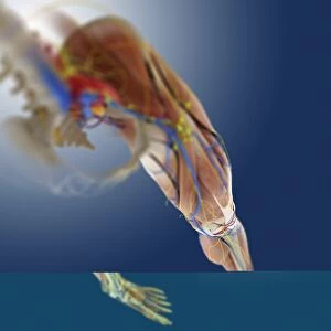
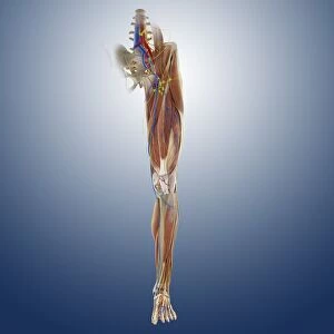
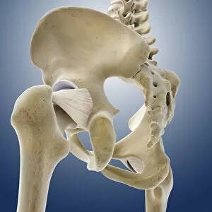
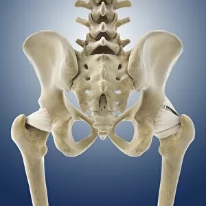
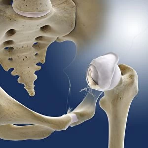
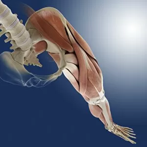
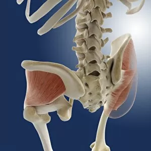
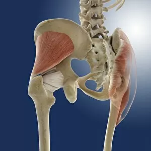
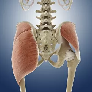
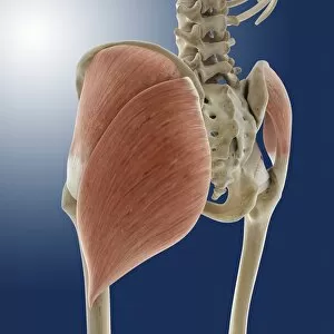
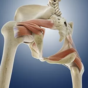
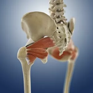
The hip bone, a vital component of the pelvis, plays a crucial role in our daily activities. In its normal anterior view, it showcases the intricate structure that supports our body's movements. However, repetitive actions like aerobics or knee-lifting exercises can lead to trauma and potential damage. Arthritis and osteophytes on the femoral heads are visible in an anterior view of the pelvis with hip bones. These conditions highlight the importance of maintaining joint health and seeking appropriate treatment when necessary. An open hip reveals its fascinating anatomy, showcasing the articular surface of the femur. This illustration provides insight into how this complex joint functions seamlessly during movement. Scientific illustrations depicting hip bone ligaments and joints offer valuable insights into human anatomy. Understanding these structures is essential for medical professionals who diagnose and treat various conditions related to this area. Buttock muscles play a significant role in supporting hip function as depicted in artwork C013/4414. Strengthening these muscles through targeted exercises can help prevent injuries and maintain overall stability. Degeneration of the hip joint known as Perthes disease is visualized through digital illustrations. This condition emphasizes the need for early detection and intervention to preserve proper joint function. Damaged hips exemplify different scenarios where injury or disease has affected this crucial skeletal structure within the human pelvis. These examples serve as reminders to prioritize orthopedic health throughout life. Artwork F007/7208 showcases detailed depictions of individual hip bones, highlighting their unique characteristics while emphasizing their interconnectedness within our bodies' framework. Female pelvic anatomy artwork sheds light on gender-specific considerations regarding hip bone structure and functionality—a reminder that healthcare must address diverse anatomical variations among individuals effectively. Lastly, artwork F006/3083 offers another perspective on understanding hip bones—underscoring their significance from an artistic standpoint while reminding us about their biological importance in supporting our everyday movements.