
Hormonal Collection (#3)
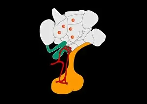
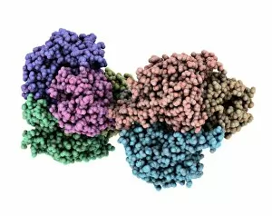
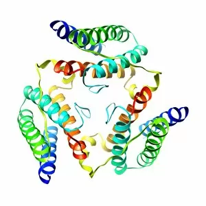
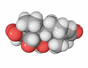
"Hormonal: Unveiling the Intricacies of our Body's Chemical Messengers" Delving into the depths of our brain, the medulla oblongata holds the key to hormonal regulation


For sale as Licensed Images
Choose your image, Select your licence and Download the media
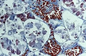
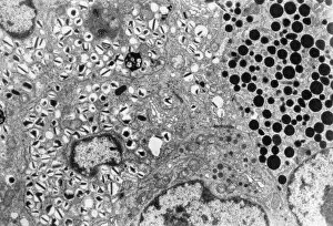
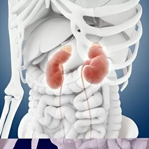
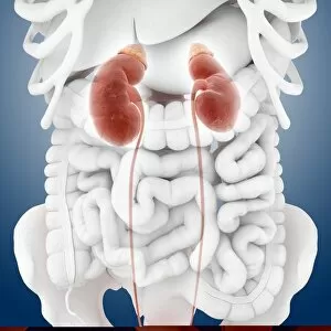
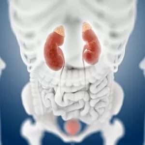
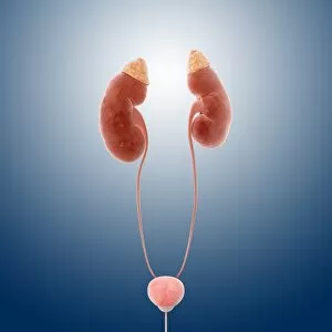
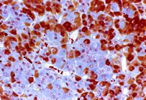
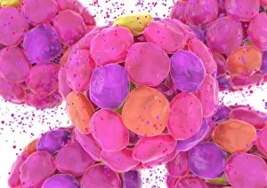
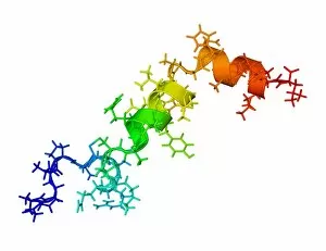
"Hormonal: Unveiling the Intricacies of our Body's Chemical Messengers" Delving into the depths of our brain, the medulla oblongata holds the key to hormonal regulation. Behold the mesmerizing artwork capturing cortisol crystals under a light micrograph - a glimpse into stress management at its core. Explore the intricate anatomy of the pancreas through captivating artwork, where hormones play their symphony. Witness the beauty within Islets of Langerhans under a light micrograph, revealing their crucial role in blood sugar control. The oxytocin neurotransmitter molecule - an enchanting messenger that fosters love and bonding in our brains. Dive into an artistic representation of thyroid anatomy, unraveling its significance in metabolism and energy balance. Marvel at SEM images showcasing thyroid gland capillaries and blood vessels - lifelines for hormone distribution throughout our body. Journey through the brain's limbic system, where emotions intertwine with hormonal signals to shape our experiences. Admire exquisite artwork depicting Islets of Langerhans cells - guardians ensuring harmony within our endocrine system. Embark on an anatomical exploration of kidney function through stunning artwork, highlighting its role in hormone production and filtration processes. In this captivating journey, we uncover how these hormonal cues orchestrate countless bodily functions with precision and grace – truly a testament to nature's brilliance.