
Host Cell Collection
"Exploring the Intricate World of Host Cells: Unveiling the Battles Within" In this captivating journey, we delve into the fascinating realm of host cells


For sale as Licensed Images
Choose your image, Select your licence and Download the media
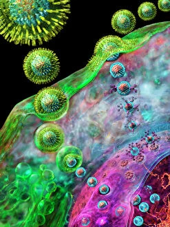
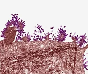
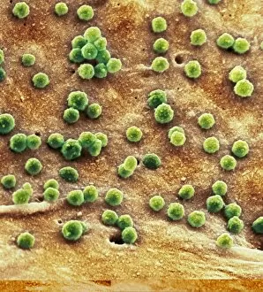
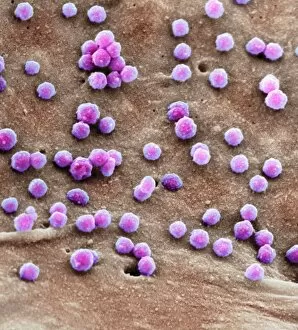
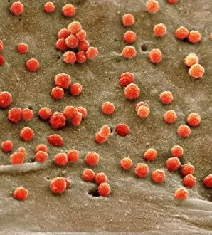
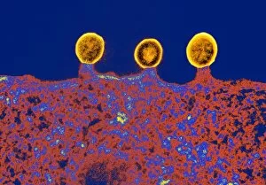
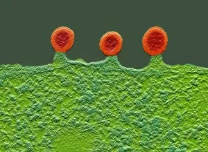
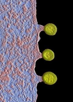
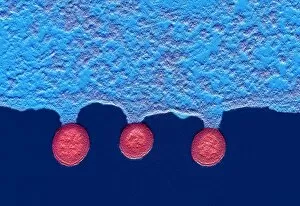
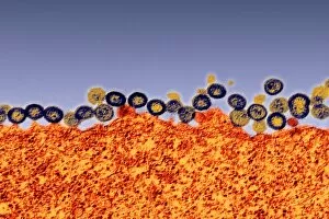
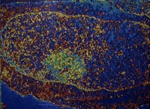
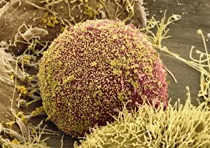
"Exploring the Intricate World of Host Cells: Unveiling the Battles Within" In this captivating journey, we delve into the fascinating realm of host cells, where life-altering battles take place. Witness as the Herpes virus replicates within its unsuspecting host, leaving a trail of havoc in its wake. Marvel at the resilience of Simian immunodeficiency virus (SIV), cunningly evading detection while silently compromising immune systems. As our exploration continues, we encounter Chlamydia infection portrayed through stunning artwork C016 / 8952, C016 / 8954, and C016 / 8953. These visual representations offer glimpses into the intricate dance between pathogen and host cell – an ongoing struggle for dominance. The Flu virus emerges next under a powerful microscope's lens – its TEM image revealing its spherical structure with menacing spikes. This tiny entity wreaks havoc on millions worldwide each year. But perhaps one of the most notorious adversaries is HIV - Human Immunodeficiency Virus. Through SEM images such as C014 / 0581, C014 / 0580, C014 / 0579, C017 / 8338, C017/8339 andC017/8337; we witness infected cells battling against this relentless invader that hijacks their machinery to replicate itself relentlessly. These snapshots remind us that within every seemingly ordinary cell lies an epic struggle for survival against these invisible foes. The complex interactions between pathogens and their hosts continue to captivate scientists worldwide as they strive to unravel nature's secrets. So let us marvel at these microscopic battlegrounds where viruses invade and exploit their unwitting hosts' cellular machinery. In understanding these encounters better lies hope for developing novel treatments or even preventing future outbreaks altogether—shedding light on a world unseen yet profoundly impactful on human health.