
Human Small Intestine Collection
The human small intestine is a vital component of the digestive system, responsible for breaking down food and absorbing nutrients into the bloodstream


For sale as Licensed Images
Choose your image, Select your licence and Download the media
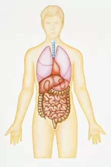
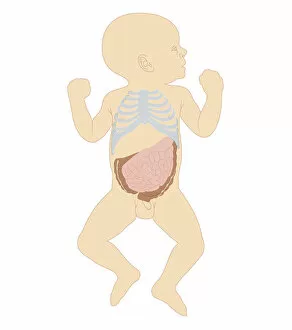
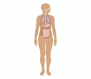
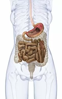
The human small intestine is a vital component of the digestive system, responsible for breaking down food and absorbing nutrients into the bloodstream. This intricate organ can be visualized through various biomedical illustrations. One such illustration showcases a cross section of the human digestive system connected to the esophagus. It provides a detailed view of how food travels through this complex network, passing through the stomach and eventually reaching the small intestine. Another diagram focuses on the abdominal cavity in the human body, with an emphasis on the small intestine. The liver has been removed to highlight this crucial organ's location and function within our bodies. Additionally, an illustration depicts both the digestive and respiratory systems, emphasizing their interconnectedness. This serves as a reminder that oxygen from respiration fuels digestion in order to sustain life. Furthermore, cross-sectional biomedical illustrations offer insights into specific demographics. For instance, one image displays a newborn baby boy's intestines alongside his rib cage - highlighting their delicate nature at such an early stage of development. Similarly, another cross-section reveals differences between genders by showcasing a girl's digestive system. These visuals help us understand variations in anatomy while appreciating our shared biological processes. Chemical breakdown within our digestive system is also illustrated meticulously; it demonstrates how enzymes break down food particles into smaller components that can be absorbed by intestinal villi for further processing or excretion. Moreover, these biomedical illustrations provide comprehensive views of not only individual organs but also their placement within our bodies as seen in cross-sectional depictions of large and small intestines along with pelvis structures in adult females. In addition to static images, dynamic procedures like colonoscopy are captured via illustrative visuals. Such imagery aids medical professionals during diagnostic examinations while offering valuable insights into potential health issues affecting adults specifically female patients' intestinal tracts. Lastly, microscopic views reveal intimate details about duodenal villi found within our intestines – tiny finger-like projections responsible for nutrient absorption. By magnifying these structures, scientists can better understand their role in maintaining our overall health.