
Icosahedral Collection
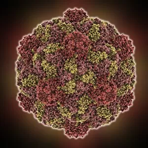
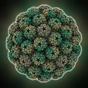
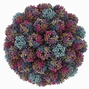
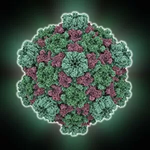
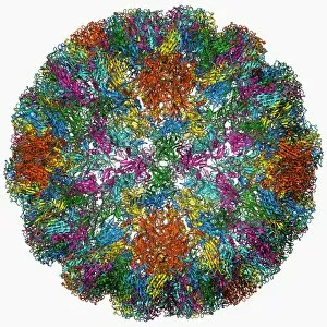
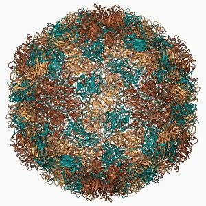
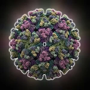
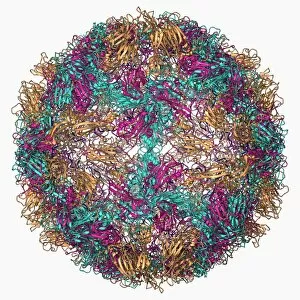
"Icosahedral: Unveiling the Intricate World of Viral Architecture" Delving into the microscopic realm


For sale as Licensed Images
Choose your image, Select your licence and Download the media
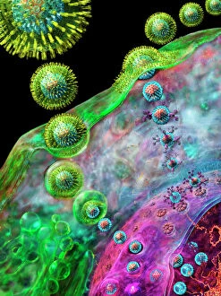
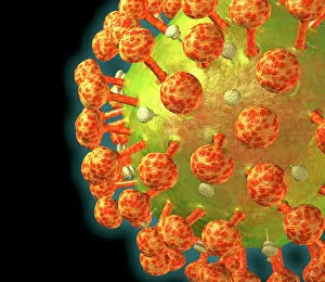
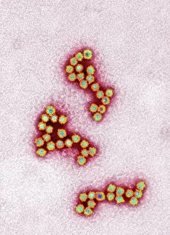
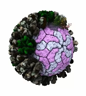
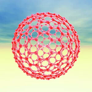
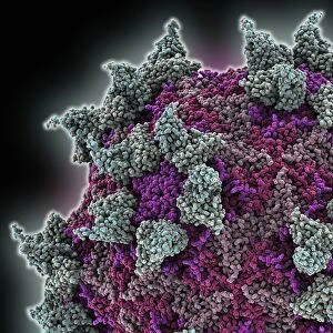
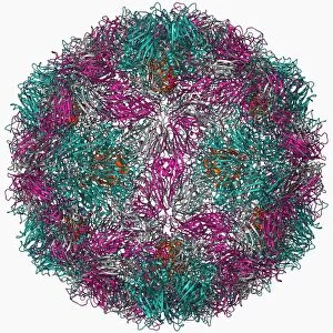
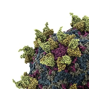
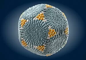
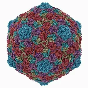
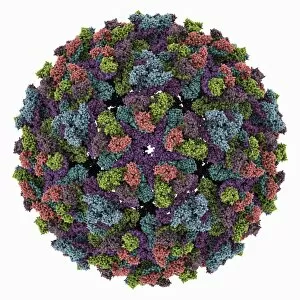
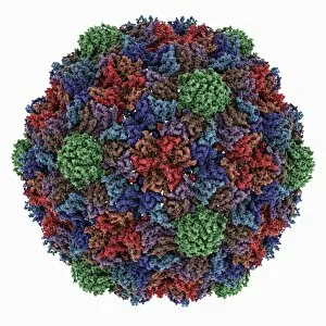
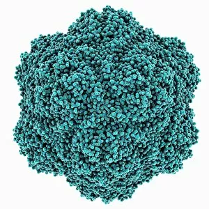
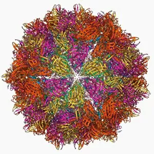
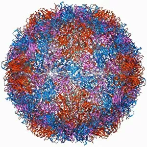
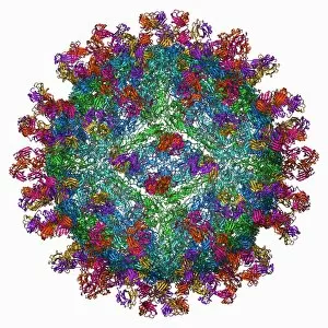
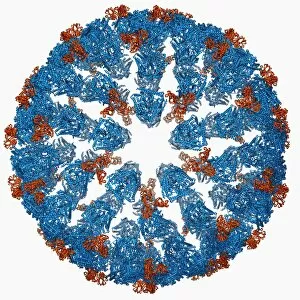
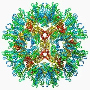
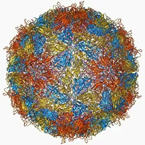
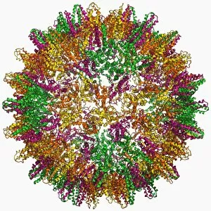
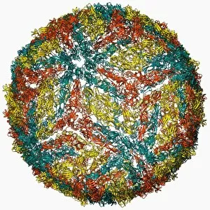
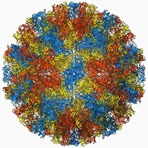
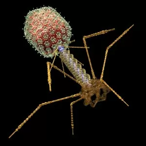
"Icosahedral: Unveiling the Intricate World of Viral Architecture" Delving into the microscopic realm, we encounter the mesmerizing icosahedral structure of an HIV particle, a key player in the battle against AIDS. Norovirus particles reveal their symmetrical beauty under the watchful eye of a Transmission Electron Microscope (TEM), showcasing nature's intricate design. Witnessing the replication process, we observe Herpes virus multiplying within host cells, unraveling its secrets through captivating imagery. Through computer artwork, Herpes virus particles come to life with stunning detail and precision, offering us a glimpse into their complex composition. The artistic representation of a Rotavirus particle takes center stage as it showcases its unique icosahedral shape that contributes to its infectious potency. Exploring beyond viruses, we delve into the world of molecules with computer-generated artwork depicting Fullerene - an iconic molecule boasting an icosahedral framework. A virtual journey inside herpes-infected cells reveals intricately designed computer artwork capturing individual herpes virus particles in all their glory. With meticulous attention to detail, computer-generated artistry unveils multiple perspectives of herpes virus particles - each one showcasing their distinctive features and allure. Adenovirus particles take on tangible form through vivid illustrations that highlight their remarkable icosahedral symmetry and structural elegance. In molecular model C015 / 7139, Rhinovirus joins forces with antibodies in an intricate dance – revealing how our immune system combats this common cold culprit at a molecular level. Molecular model F006 / 9431 brings forth Rhinovirus' capsid structure in breathtaking clarity – unlocking new insights into this notorious viral family responsible for countless sniffles worldwide. Further exploring Rhinovirus interactions at a molecular scale is showcased by molecular model C015 / 7138, where antibodies stand as our allies against this relentless respiratory invader.