
Ileum Collection (#2)

The ileum, a vital part of the human digestive system, plays a crucial role in nutrient absorption

Background imageIleum Collection: Illustration showing human digestive system

Illustration showing human digestive system
Medicine: Human anatomy, digestive system. Drawing

Background imageIleum Collection: Human intestines, historical model

Human intestines, historical model
Human intestines. 1940s anatomical model of the human gut, showing the small intestine at centre (pink), with the large intestine (grey) around it

Background imageIleum Collection: Ileocaecal valve, endoscopic view C016 / 8317

Ileocaecal valve, endoscopic view C016 / 8317
Ileocaecal valve. Endoscopic view of the healthy interior of a persons ileum (small intestine), showing the ileocaecal valve

Background imageIleum Collection: Diseased organs, artwork

Diseased organs, artwork
Diseased organs. Computer artwork showing diseased major organs of the human torso. From top to bottom: larynx (voicebox), trachea (windpipe), lungs (centre left and right), heart (centre), liver

Background imageIleum Collection: Intestinal tumour, artwork C015 / 6451

Intestinal tumour, artwork C015 / 6451
Intestinal tumour. Computer artwork of a tumour on the internal wall of an intestine

Background imageIleum Collection: Digestive system, artwork C015 / 6450

Digestive system, artwork C015 / 6450
Human anatomy. Computer artwork showing the organs of the human digestive system and their relative positions within the body

Background imageIleum Collection: Male anatomy, artwork C015 / 6448

Male anatomy, artwork C015 / 6448
Male anatomy. Computer artwork showing the major organs of the human torso. From top to bottom: oesophagus (white, top centre), trachea (windpipe, pink, top centre)

Background imageIleum Collection: Female anatomy, artwork C015 / 6447

Female anatomy, artwork C015 / 6447
Female anatomy. Computer artwork showing the major organs of the femaletorso. From top to bottom: oesophagus (white, top centre), trachea (windpipe, pink, top centre)

Background imageIleum Collection: Food & bacteria in the ileum, SEM

Food & bacteria in the ileum, SEM
Food and bacteria in the small intestine. Coloured scanning electron micrograph (SEM) of food debris (yellow) and normal intestinal bacteria (purple) on villi (projections, red) in the ileum

Background imageIleum Collection: Small intestine, barium X-ray

Small intestine, barium X-ray
Small intestines, coloured X-ray. The four X-rays reveal the passage of a swallowed barium contrast medium through the upper part of the digestive system. The medium shows up white on X-rays

Background imageIleum Collection: Coloured SEM of a sectioned villus from the ileum

Coloured SEM of a sectioned villus from the ileum

Background imageIleum Collection: Irritable bowel syndrome

Irritable bowel syndrome. Artwork of a digestive system suffering from irritable bowel syndrome (IBS). IBS is also known as spastic colon or irritable colon syndrome

Background imageIleum Collection: Crohns disease, X-ray

Crohns disease, X-ray

Background imageIleum Collection: Illustration of Crohns disease of the ileum

Illustration of Crohns disease of the ileum

Background imageIleum Collection: Peyers patches, SEM

Peyers patches, SEM
Peyers patches (red), coloured scanning electron micrograph (SEM). These regions of lymphoid tissue are found in the human intestine, usually in the ileum, the final section of the small intestine

Background imageIleum Collection: Small intestine section, light micrograph

Small intestine section, light micrograph
Small intestine section. Light micrograph of a vertical section through the illeum, part of the small intestine. The upper layer (the digestive surface)

Background imageIleum Collection: Small intestine lining, light micrograph

Small intestine lining, light micrograph
Small intestine lining. Light micrograph of a section through the finger-like projections (villi) of the duodenum, the uppermost part of the small intestine

Background imageIleum Collection: Locust internal anatomy, light micrograph

Locust internal anatomy, light micrograph
Locust internal anatomy. Light micrograph of a transverse section through the gut and part of the male reproductive system of the locust


For sale as Licensed Images

Choose your image, Select your licence and Download the media

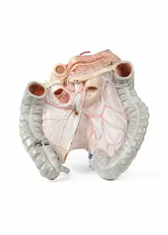
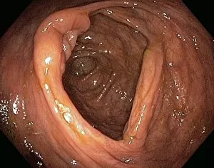
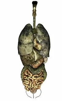
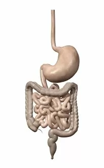
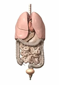
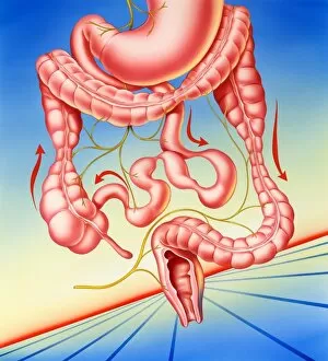
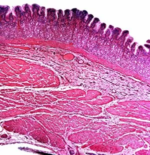
The ileum, a vital part of the human digestive system, plays a crucial role in nutrient absorption. Located in the small intestine, it is responsible for breaking down food particles and extracting essential nutrients to nourish our body. In a cross-sectional biomedical illustration of the human digestive system connected to the esophagus, we can see how the ileum forms an integral part of this intricate network. Its inner lining is designed with tiny finger-like projections called villi that increase its surface area for maximum nutrient absorption. A female body showcasing both the digestive and circulatory systems highlights how these two systems work hand in hand to ensure proper functioning of our body. The ileum's close proximity to blood vessels allows for efficient transport of absorbed nutrients throughout our bloodstream. Similarly, an anatomy depiction of male respiratory organs and internal organs emphasizes how interconnected our bodily systems truly are. The ileum works alongside other organs such as the lungs and liver to maintain overall health and well-being. Visualizing internal organ structures through 3D renderings provides us with a deeper understanding of their complexity. A healthy female's internal organs exhibit remarkable intricacy, including the ileum nestled within her digestive system. Meanwhile, a male anatomy diagram showcases various internal organs against a blue background - illustrating their location within his body while highlighting the importance of each individual component. Examining front view illustrations helps us comprehend how different parts come together harmoniously within our bodies' complex machinery. Observing an anatomical representation from this perspective reveals where exactly we can find the ileum amidst other components like stomach or intestines. Superimposing interior organ images onto a woman's midsection further enhances our comprehension by providing visual context on their placement within her body structure. This technique aids in recognizing precisely where one would locate vital elements like the ileum when examining real-life scenarios or medical cases. Lastly, exploring general colon anatomy with labels gives us insight into specific regions along our intestinal tract.