
Immunoglobulin Collection
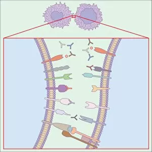
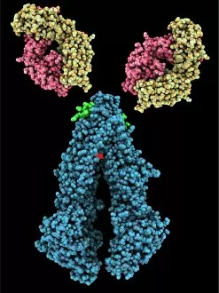
"Unleashing the Power of Immunoglobulin: Unveiling the Mighty Defenders" Immunoglobulins, also known as antibodies


For sale as Licensed Images
Choose your image, Select your licence and Download the media
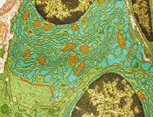
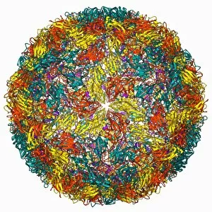
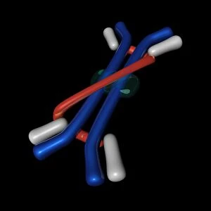
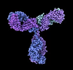
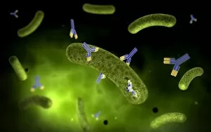
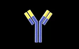
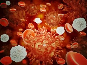
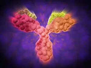
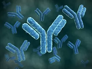
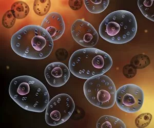
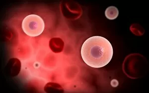
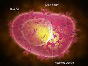
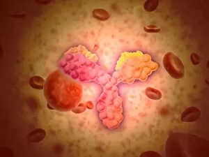
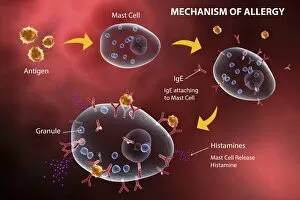
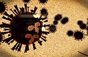
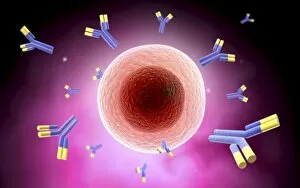
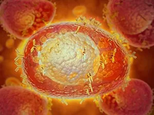
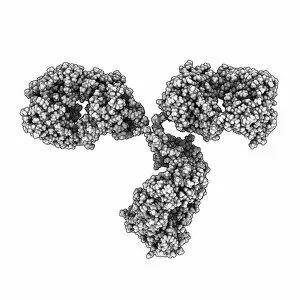
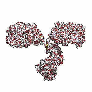
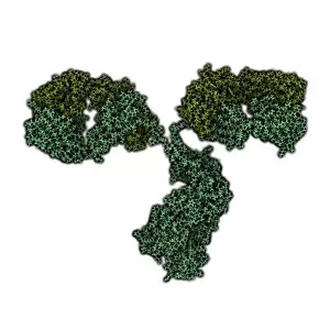
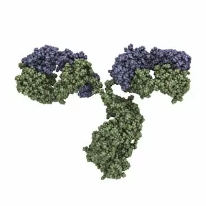
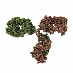
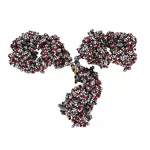
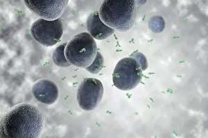
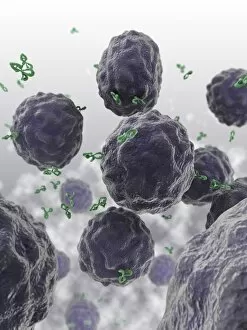
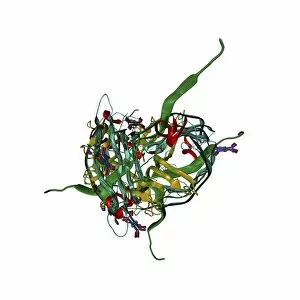
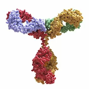
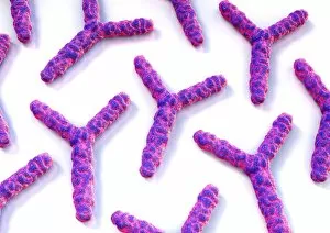
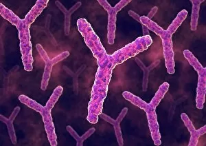
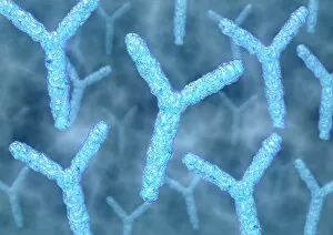
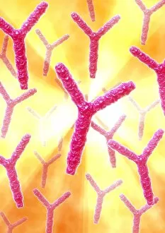
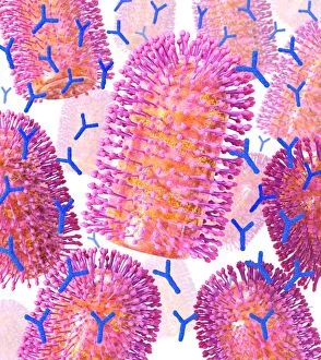
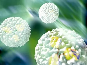
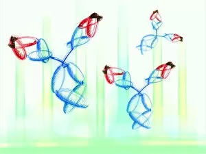
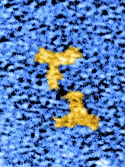
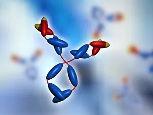
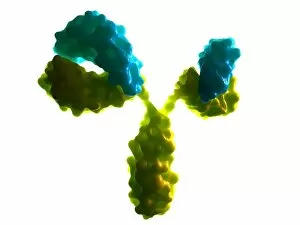
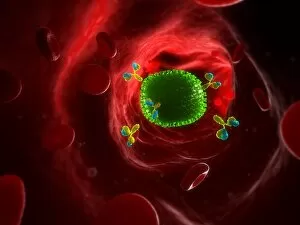
"Unleashing the Power of Immunoglobulin: Unveiling the Mighty Defenders" Immunoglobulins, also known as antibodies, are remarkable proteins produced by plasma cells in our immune system. These tiny warriors play a crucial role in safeguarding our health against various pathogens and diseases. In this captivating image captured through Transmission Electron Microscopy (TEM), we witness the Foot-and-mouth disease virus F006 / 9556 being targeted by an Immunoglobulin G antibody molecule C016 / 4462. This conceptual representation showcases how antibodies attach to harmful bacteria, effectively neutralizing and eliminating them. Zooming into the microscopic world of human B-cells, we observe their intricate structure responsible for producing these powerful immunoglobulins. Their tireless efforts ensure that our bodies are equipped with a diverse arsenal of defense mechanisms. Another conceptual image portrays an antibody attaching itself to a bacterium, illustrating its ability to recognize and eliminate specific threats with precision and efficiency. This visual depiction highlights the extraordinary specificity exhibited by immunoglobulins. Further exploration reveals microscopic views of human antibodies interacting with red blood cells – a testament to their constant vigilance in protecting us from harm. The complexity and elegance displayed in these images emphasize the intricate dance between our immune system's components. Not limited to bacterial foes alone, immunoglobulins also combat viral intruders like H5N1 virus alongside red blood cells and white blood cells as seen under high magnification microscopy. Witnessing this battle on such a minuscule scale reminds us of the relentless fight occurring within our bodies every day. Additionally, we encounter natural killer cells injecting toxins into bacteria - yet another awe-inspiring display of immunity at work. These specialized cells act swiftly against invaders, reinforcing our defenses when needed most.