
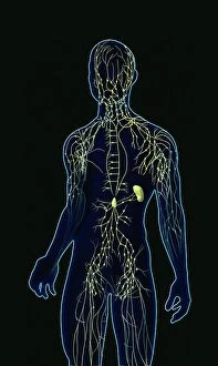
Immunological Collection (#2)
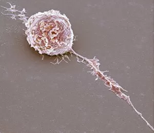
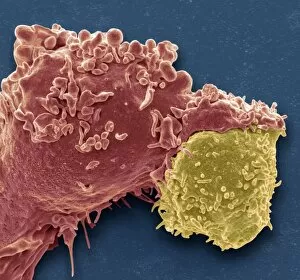
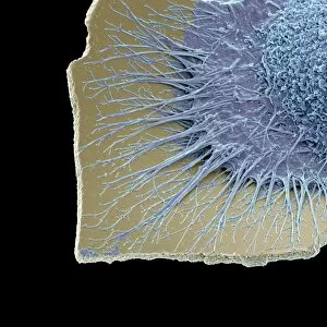
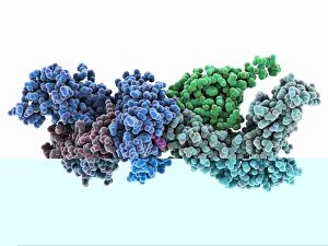
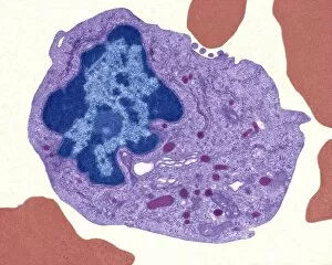
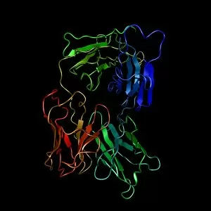
"Unlocking the Power of Immunological Defense: A Visual Journey" Witness the incredible battle between a Neutrophil and MRSA as it engulfs the harmful bacteria


For sale as Licensed Images
Choose your image, Select your licence and Download the media
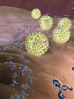
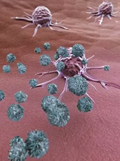
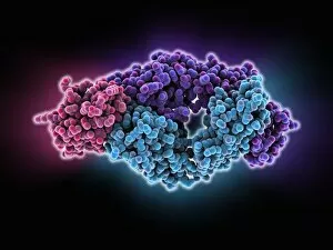
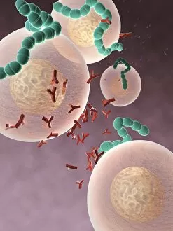
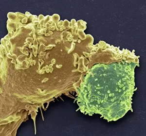
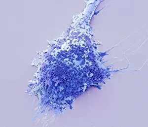
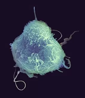
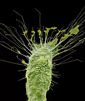
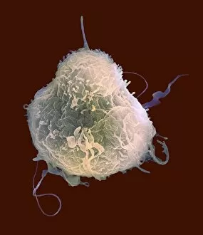
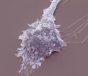
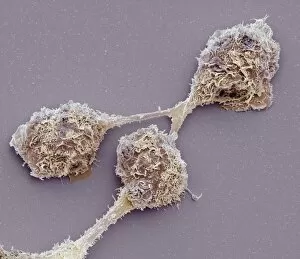
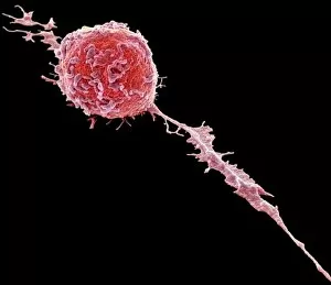
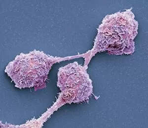
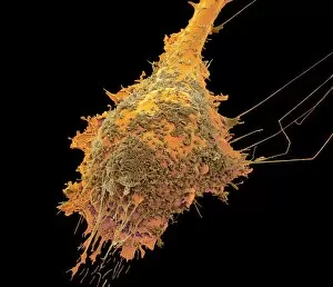
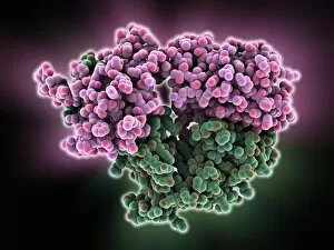
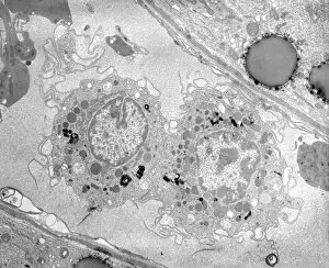
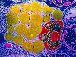
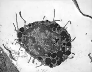
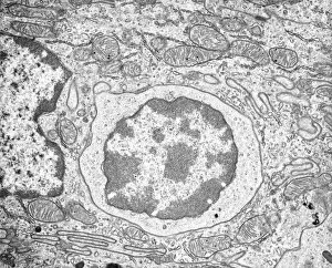
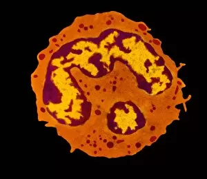
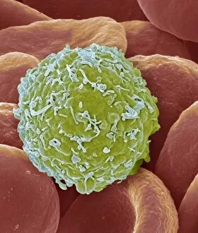
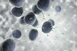
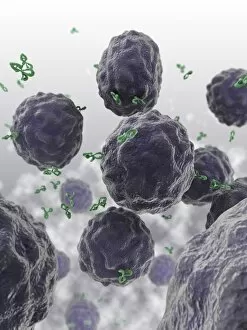
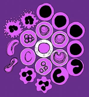
"Unlocking the Power of Immunological Defense: A Visual Journey" Witness the incredible battle between a Neutrophil and MRSA as it engulfs the harmful bacteria, captured in stunning detail through SEM C018 / 8596. Delve into the intricate world of phagocytosis as fungal spores are devoured by specialized cells, showcased beautifully in an SEM image. Behold the might of an Activated Macrophage, ready to defend our bodies against invading pathogens, magnified through SEM C015 / 6375. Explore the artistic representation of Lymphocyte white blood cells, showcasing their crucial role in immune response with captivating artwork. Marvel at Dendritic cells depicted in breathtaking artwork - these sentinel-like cells play a pivotal role in initiating immune responses. Peer into the microscopic realm with TEM imagery revealing Macrophage cells at work, tirelessly engulfing foreign invaders to protect us from harm. Immerse yourself in an illustration depicting White and red blood cells working harmoniously to maintain our body's defense mechanisms intact. Observe another mesmerizing SEM image capturing Phagocytosis of fungus spores - witness how our immune system eliminates potential threats. Discover the power of Immunoglobulin G antibody alongside egg white F006 / 9682 - highlighting how antibodies neutralize harmful substances within our bodies. Uncover a TEM snapshot showcasing Macrophages and lymphocytes collaborating seamlessly to mount a formidable immunological defense against intruders. Dive deeper into immunity with striking SEM C016 / 3099 imagery displaying White blood cells and platelets orchestrating their protective roles within our bloodstream. Experience another perspective on this dynamic duo as they come together under SEM C016 / 3098 - marvel at their collective strength safeguarding our well-being.