
Infect Collection
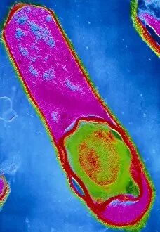
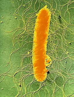
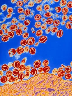
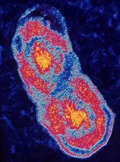
"Infect: Unseen Threats and Devastating Consequences" Coloured TEM of a Salmonella bacterium: Revealing the microscopic world


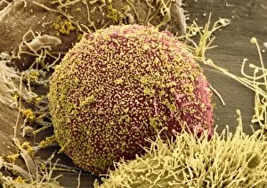
For sale as Licensed Images
Choose your image, Select your licence and Download the media
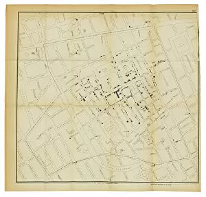
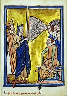
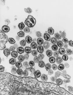
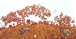
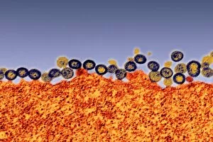
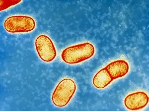
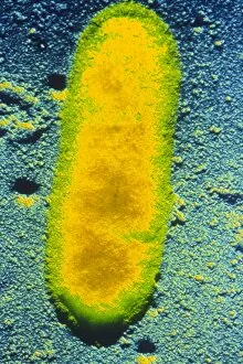
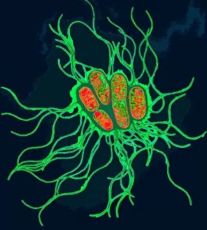
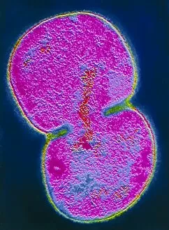
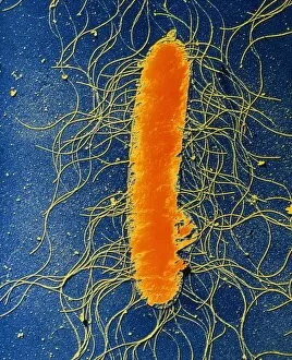
"Infect: Unseen Threats and Devastating Consequences" Coloured TEM of a Salmonella bacterium: Revealing the microscopic world, this image captures the vibrant colors of a Salmonella bacterium, reminding us that danger can lurk where we least expect it. A map from On the Mode of Communication of Cholera, 1855 (litho): This historical map serves as a haunting reminder of how diseases like cholera can rapidly spread through communities, emphasizing the importance of understanding and preventing infections. The fourth plague of Egypt, from a book of Bible Pictures, c. 1250 (vellum): Depicting one of the biblical plagues sent to punish Pharaoh for his refusal to release the Israelites, this illustration showcases how infectious diseases have plagued humanity since ancient times. Common Ash dieback caused by Ash Dieback fungal disease: Witnessing nature's vulnerability to infection, this image portrays ash trees suffering from Chalara fraxinea fungus—a stark reminder that even our environment is not immune to devastating contagions. TEM of HIV (AIDS) viruses budding from a T-cell: Offering insight into one of modern medicine's greatest challenges, this electron micrograph highlights HIV/AIDS viruses emerging from T-cells—an ongoing battle against an insidious infection. Proteus mirabilis bacterium: Capturing the intricate details underpinning microbial lifeforms, this photograph showcases Proteus mirabilis bacteria—a constant reminder that unseen pathogens are ever-present in our surroundings. Chalara dieback sign in woodland with infected ash trees: Standing amidst an afflicted forest landscape ravaged by highly contagious fungal disease, Lower Wood Reserve reminds us that infections can devastate entire ecosystems if left unchecked. 8 & Dairy farming heifers with Ringworm infection.