Internal Anatomy Collection (#19)

"Exploring the Intricacies: Unveiling Internal Anatomy through Art and Science" Delving into the hidden world of internal anatomy

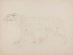
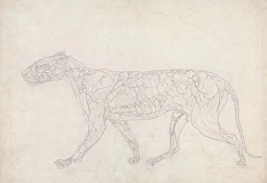
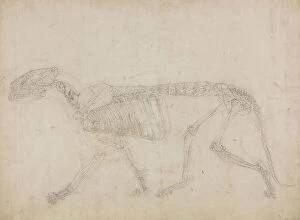
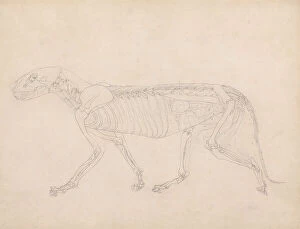
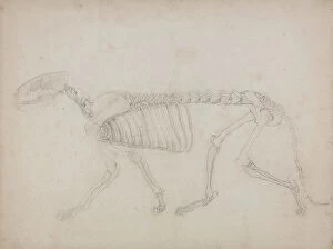
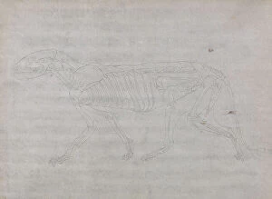
Background imageInternal Anatomy Collection: Study of a Tiger, Lateral View, from A Comparative Anatomical Exposition of the Structure of

Study of a Tiger, Lateral View, from A Comparative Anatomical Exposition of the Structure of the Human Body with that
XYC261343 Study of a Tiger, Lateral View, from A Comparative Anatomical Exposition of the Structure of the Human Body with that of a Tiger and a Common Fowl

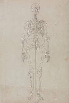
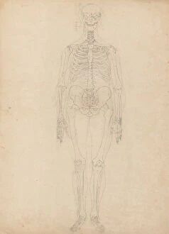
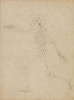
Background imageInternal Anatomy Collection: Study of the Human Figure, Anterior View, from A Comparative Anatomical Exposition of

Study of the Human Figure, Anterior View, from A Comparative Anatomical Exposition of the Structure of the Human Body
XYC257409 Study of the Human Figure, Anterior View, from A Comparative Anatomical Exposition of the Structure of the Human Body with that of a Tiger and a Common Fowl

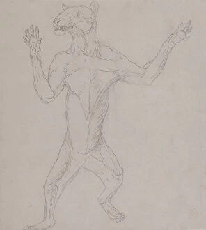
Background imageInternal Anatomy Collection: Study of a Tiger, Standing in Human Posture, from A Comparative Anatomical Exposition of

Study of a Tiger, Standing in Human Posture, from A Comparative Anatomical Exposition of the Structure of the Human
XYC261362 Study of a Tiger, Standing in Human Posture, from A Comparative Anatomical Exposition of the Structure of the Human Body with that of a Tiger and a Common Fowl

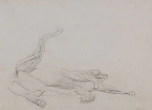
Background imageInternal Anatomy Collection: Study of a Tiger, Recumbent, Viewed Ventrally, from A Comparative Anatomical Exposition of

Study of a Tiger, Recumbent, Viewed Ventrally, from A Comparative Anatomical Exposition of the Structure of the Human
XYC261361 Study of a Tiger, Recumbent, Viewed Ventrally, from A Comparative Anatomical Exposition of the Structure of the Human Body with that of a Tiger and a Common Fowl

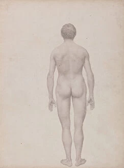
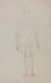
Background imageInternal Anatomy Collection: Study of the Human Figure, Posterior View, Undissected, Finished Study for Table VII

Study of the Human Figure, Posterior View, Undissected, Finished Study for Table VII
XYC261316 Study of the Human Figure, Posterior View, Undissected, Finished Study for Table VII, from A Comparative Anatomical Exposition of the Structure of the Human Body with that of a Tiger

Background imageInternal Anatomy Collection: Study of the Human Figure, Anterior View, Diagram for Key to Table I

Study of the Human Figure, Anterior View, Diagram for Key to Table I, from A Comparative Anatomical Exposition of
XYC261303 Study of the Human Figure, Anterior View, Diagram for Key to Table I, from A Comparative Anatomical Exposition of the Structure of the Human Body with that of a Tiger and a Common Fowl

Background imageInternal Anatomy Collection: Organs of intestinal absorption - The normal life & health of J. Rengade. Drawing by A. Demarle 1881

Organs of intestinal absorption - The normal life & health of J. Rengade. Drawing by A. Demarle 1881
JAB4120077 Organs of intestinal absorption - The normal life & health of J. Rengade. Drawing by A. Demarle 1881 by Demarle

Background imageInternal Anatomy Collection: The child in the breast (J. Rengade's normal life & health) - Breastfeeding. Drawing by A

The child in the breast (J. Rengade's normal life & health) - Breastfeeding. Drawing by A. Demarle 1881
JAB4120219 The child in the breast (J. Rengade's normal life & health) - Breastfeeding. Drawing by A. Demarle 1881 by Demarle, Alphonse (1826-1896); (add.info.: The child in the breast (J)

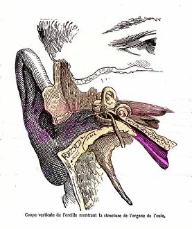
Background imageInternal Anatomy Collection: Ear cut, hearing - The normal life and health of J. Rengade. Drawing A. Demarle. 1881

Ear cut, hearing - The normal life and health of J. Rengade. Drawing A. Demarle. 1881
JAB4120139 Ear cut, hearing - The normal life and health of J. Rengade. Drawing A. Demarle. 1881 by Demarle, Alphonse (1826-1896); (add.info.: Ear cut, hearing - The normal life and health of J)

Background imageInternal Anatomy Collection: Cranial nerves - The normal life and health of J. Rengade-Drawing A. Demarle 1881

Cranial nerves - The normal life and health of J. Rengade-Drawing A. Demarle 1881
JAB4120319 Cranial nerves - The normal life and health of J. Rengade-Drawing A. Demarle 1881 by Demarle, Alphonse (1826-1896); (add.info.: Cranial nerves - The normal life and health of J)

Background imageInternal Anatomy Collection: L'encephalus (The normal life & health of J. Rengade) - Fig du Top: profile view

L'encephalus (The normal life & health of J. Rengade) - Fig du Top: profile view. Fig 2: Vertical cut.Drawing A
JAB4120241 L'encephalus (The normal life & health of J. Rengade) - Fig du Top: profile view. Fig 2: Vertical cut.Drawing A

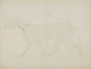
Background imageInternal Anatomy Collection: Study of a Tiger, Lateral View, Diagram Showing the Subcutaneous Blood Supply

Study of a Tiger, Lateral View, Diagram Showing the Subcutaneous Blood Supply
XYC261342 Study of a Tiger, Lateral View, Diagram Showing the Subcutaneous Blood Supply, from A Comparative Anatomical Exposition of the Structure of the Human Body with that of a Tiger

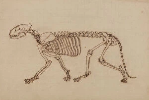
Background imageInternal Anatomy Collection: Study of a Tiger, Lateral View, from A Comparative Anatomical Exposition of the Structure of

Study of a Tiger, Lateral View, from A Comparative Anatomical Exposition of the Structure of the Human Body with that
XYC261340 Study of a Tiger, Lateral View, from A Comparative Anatomical Exposition of the Structure of the Human Body with that of a Tiger and a Common Fowl

Background imageInternal Anatomy Collection: Study of a Fowl, Lateral View, Diagram for Key to Table XV, from A Comparative Anatomical

Study of a Fowl, Lateral View, Diagram for Key to Table XV, from A Comparative Anatomical Exposition of the Structure
XYC261310 Study of a Fowl, Lateral View, Diagram for Key to Table XV, from A Comparative Anatomical Exposition of the Structure of the Human Body with that of a Tiger and a Common Fowl

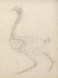
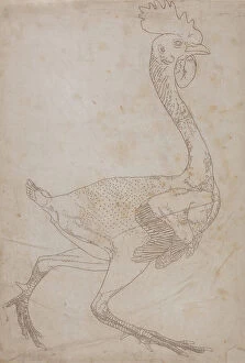
Background imageInternal Anatomy Collection: Study of a Fowl, Lateral View, from A Comparative Anatomical Exposition of the Structure of

Study of a Fowl, Lateral View, from A Comparative Anatomical Exposition of the Structure of the Human Body with that of
XYC261368 Study of a Fowl, Lateral View, from A Comparative Anatomical Exposition of the Structure of the Human Body with that of a Tiger and a Common Fowl

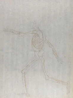
Background imageInternal Anatomy Collection: Study of the Human Figure, Lateral View, from A Comparative Anatomical Exposition of the Structure

Study of the Human Figure, Lateral View, from A Comparative Anatomical Exposition of the Structure of the Human Body
XYC261323 Study of the Human Figure, Lateral View, from A Comparative Anatomical Exposition of the Structure of the Human Body with that of a Tiger and a Common Fowl

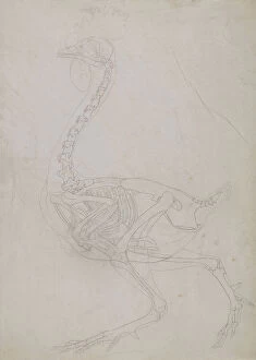
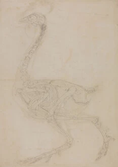
Background imageInternal Anatomy Collection: Study of a Fowl, Lateral View, from A Comparative Anatomical Exposition of the Structure of

Study of a Fowl, Lateral View, from A Comparative Anatomical Exposition of the Structure of the Human Body with that of
XYC262888 Study of a Fowl, Lateral View, from A Comparative Anatomical Exposition of the Structure of the Human Body with that of a Tiger and a Common Fowl

Background imageInternal Anatomy Collection: Study of a Tiger, Lateral View, from A Comparative Anatomical Exposition of the Structure of

Study of a Tiger, Lateral View, from A Comparative Anatomical Exposition of the Structure of the Human Body with that
XYC261339 Study of a Tiger, Lateral View, from A Comparative Anatomical Exposition of the Structure of the Human Body with that of a Tiger and a Common Fowl

Background imageInternal Anatomy Collection: Study of a Tiger, Lateral View, Abdominal Viscera Exposed, from A Comparative Anatomical

Study of a Tiger, Lateral View, Abdominal Viscera Exposed, from A Comparative Anatomical Exposition of the Structure of
XYC261309 Study of a Tiger, Lateral View, Abdominal Viscera Exposed, from A Comparative Anatomical Exposition of the Structure of the Human Body with that of a Tiger and a Common Fowl

Background imageInternal Anatomy Collection: Study of a Fowl, Lateral View, from A Comparative Anatomical Exposition of the Structure of

Study of a Fowl, Lateral View, from A Comparative Anatomical Exposition of the Structure of the Human Body with that of
XYC261366 Study of a Fowl, Lateral View, from A Comparative Anatomical Exposition of the Structure of the Human Body with that of a Tiger and a Common Fowl

Background imageInternal Anatomy Collection: Study of a Tiger, Lateral View, from A Comparative Anatomical Exposition of the Structure of

Study of a Tiger, Lateral View, from A Comparative Anatomical Exposition of the Structure of the Human Body with that
XYC261337 Study of a Tiger, Lateral View, from A Comparative Anatomical Exposition of the Structure of the Human Body with that of a Tiger and a Common Fowl

Background imageInternal Anatomy Collection: Study of the Human Figure, Posterior View, from A Comparative Anatomical Exposition of

Study of the Human Figure, Posterior View, from A Comparative Anatomical Exposition of the Structure of the Human Body
XYC257414 Study of the Human Figure, Posterior View, from A Comparative Anatomical Exposition of the Structure of the Human Body with that of a Tiger and a Common Fowl

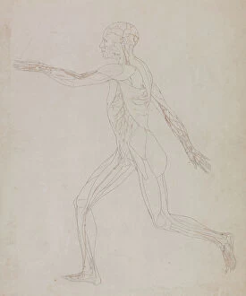
Background imageInternal Anatomy Collection: Study of the Human Figure, Lateral View, from A Comparative Anatomical Exposition of the Structure

Study of the Human Figure, Lateral View, from A Comparative Anatomical Exposition of the Structure of the Human Body
XYC261321 Study of the Human Figure, Lateral View, from A Comparative Anatomical Exposition of the Structure of the Human Body with that of a Tiger and a Common Fowl

Background imageInternal Anatomy Collection: Study of a Fowl, Lateral View, with skin and underlying fascial layers removed

Study of a Fowl, Lateral View, with skin and underlying fascial layers removed
XYC261317 Study of a Fowl, Lateral View, with skin and underlying fascial layers removed, from A Comparative Anatomical Exposition of the Structure of the Human Body with that of a Tiger

Background imageInternal Anatomy Collection: Study of the Human Figure, Lateral View, from A Comparative Anatomical Exposition of the Structure

Study of the Human Figure, Lateral View, from A Comparative Anatomical Exposition of the Structure of the Human Body
XYC257421 Study of the Human Figure, Lateral View, from A Comparative Anatomical Exposition of the Structure of the Human Body with that of a Tiger and a Common Fowl

Background imageInternal Anatomy Collection: Study of a Fowl, Lateral View, from A Comparative Anatomical Exposition of the Structure of

Study of a Fowl, Lateral View, from A Comparative Anatomical Exposition of the Structure of the Human Body with that of
XYC261365 Study of a Fowl, Lateral View, from A Comparative Anatomical Exposition of the Structure of the Human Body with that of a Tiger and a Common Fowl

Background imageInternal Anatomy Collection: Study of a Tiger, Lateral View, from A Comparative Anatomical Exposition of the Structure of

Study of a Tiger, Lateral View, from A Comparative Anatomical Exposition of the Structure of the Human Body with that
XYC261347 Study of a Tiger, Lateral View, from A Comparative Anatomical Exposition of the Structure of the Human Body with that of a Tiger and a Common Fowl

Background imageInternal Anatomy Collection: Study of a Tiger, Lateral View, from A Comparative Anatomical Exposition of the Structure of

Study of a Tiger, Lateral View, from A Comparative Anatomical Exposition of the Structure of the Human Body with that
XYC261344 Study of a Tiger, Lateral View, from A Comparative Anatomical Exposition of the Structure of the Human Body with that of a Tiger and a Common Fowl

Background imageInternal Anatomy Collection: Study of the Human Figure, Posterior View, with skin and underlying fascial tissues removed

Study of the Human Figure, Posterior View, with skin and underlying fascial tissues removed
XYC261318 Study of the Human Figure, Posterior View, with skin and underlying fascial tissues removed, from A Comparative Anatomical Exposition of the Structure of the Human Body with that of a Tiger

Background imageInternal Anatomy Collection: A seated academic nude with his hands clasped, c. 1723-24 (red & white chalk on light brown paper)

A seated academic nude with his hands clasped, c. 1723-24 (red & white chalk on light brown paper)
7415867 A seated academic nude with his hands clasped, c.1723-24 (red & white chalk on light brown paper) by Lemoyne, Francois (1688-1737); 30x18.1 cm; Private Collection; (add.info)

Background imageInternal Anatomy Collection: 7422629

7422629

Background imageInternal Anatomy Collection: 3673277

3673277

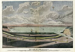
Background imageInternal Anatomy Collection: Channel Tunnel Project between Dover and Sangatte, 1906 (illustration)

Channel Tunnel Project between Dover and Sangatte, 1906 (illustration)
7415621 Channel Tunnel Project between Dover and Sangatte, 1906 (illustration) by Damblans, Eugene (1865-1945); Private Collection; (add.info.: Channel Tunnel Project between Dover (Dover)

Background imageInternal Anatomy Collection: Portrait of Giovanni Battista Bianchi, 17th century (terracotta)

Portrait of Giovanni Battista Bianchi, 17th century (terracotta)
7129493 Portrait of Giovanni Battista Bianchi, 17th century (terracotta) by Arriguzi, Fabrizio (d.1647); Palazzo Corsini, Gallerie Nazionali Barberini Corsini, Rome

Background imageInternal Anatomy Collection: Andreas Vesalius

Andreas Vesalius
3586029 Andreas Vesalius by French School, (19th century); (add.info.: Portrait of Andreas Vesalius (1514-1564) a Flemish/Netherlandish anatomist, physician, and author of books on human anatomy

Background imageInternal Anatomy Collection: Portrait of Lucrezia Panciatichi, 1541-45, (oil on wood panel)

Portrait of Lucrezia Panciatichi, 1541-45, (oil on wood panel)
7146804 Portrait of Lucrezia Panciatichi, 1541-45, (oil on wood panel) by Bronzino, Agnolo di Cosimo (1503-72); Galleria degli Uffizi, Florence, Tuscany

Background imageInternal Anatomy Collection: Two male nudes on a rock, 1890 (b/w photo)

Two male nudes on a rock, 1890 (b/w photo)
1574231 Two male nudes on a rock, 1890 (b/w photo) by Gloeden, Wilhelm von (1856-1931); (add.info.: Photograph by Wilhelm von Gloeden, (1856 - 1931): German photographer who worked mainly in Italy

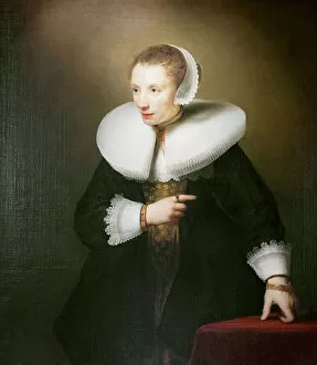
Background imageInternal Anatomy Collection: Portrait of an unknown woman, 1642-44 (oil on canvas)

Portrait of an unknown woman, 1642-44 (oil on canvas)
7146413 Portrait of an unknown woman, 1642-44 (oil on canvas) by Bol, Ferdinand (1616-80); Kenwood House, London, UK; (add.info.: Portrait of an unknown woman, 1642-44, Ferdinand Bol, oil on canvas)

Background imageInternal Anatomy Collection: Maria Bonciani, 1489 circa, (oil on panel)

Maria Bonciani, 1489 circa, (oil on panel)
7146421 Maria Bonciani, 1489 circa, (oil on panel) by Master of the Baroncelli Portraits, (c.1475-1500); Galleria degli Uffizi, Florence, Tuscany, Italy; (add.info.: Maria Bonciani, 1489 circa)

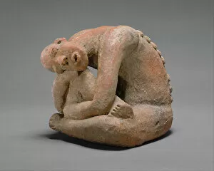
Background imageInternal Anatomy Collection: Seated Figure, 13th century (terracotta)

Seated Figure, 13th century (terracotta)
7316846 Seated Figure, 13th century (terracotta) by African School; Metropolitan Museum of Art, New York, USA; (add.info.: Middle Niger civilization.)

Background imageInternal Anatomy Collection: MELANCHTON, Philipp Schwarzerd, called (1497-1560). Theologian and Luther's collaborator (engraving)

MELANCHTON, Philipp Schwarzerd, called (1497-1560). Theologian and Luther's collaborator (engraving)
CUL4933423 MELANCHTON, Philipp Schwarzerd, called (1497-1560). Theologian and Luther's collaborator (engraving) by Durer or Duerer, Albrecht (1471-1528); (add.info.: By DURER)

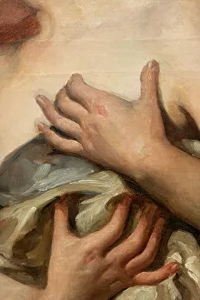
Background imageInternal Anatomy Collection: Nude Study, detail, 1878 (oil on canvas)

Nude Study, detail, 1878 (oil on canvas)
7337478 Nude Study, detail, 1878 (oil on canvas) by Agneessens, Edouard (1842-85); Musee des Beaux-Arts, Tournai, Belgium; (add.info.: Painting, Edouard Agneessens (1842-1885)

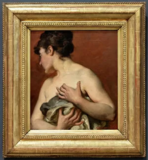
Background imageInternal Anatomy Collection: Nude Study, 1878 (oil on canvas)

Nude Study, 1878 (oil on canvas)
7337476 Nude Study, 1878 (oil on canvas) by Agneessens, Edouard (1842-85); Musee des Beaux-Arts, Tournai, Belgium; (add.info.: Painting, Edouard Agneessens (1842-1885), Nude Study (Etude de nu)

Background imageInternal Anatomy Collection: Amboise Pare

Amboise Pare
3783229 Amboise Pare; (add.info.: Ambroise Pare. French surgeon, considered as one of the fathers of surgery and modern forensic pathology; also the inventor of several surgical instruments C)

Background imageInternal Anatomy Collection: Mademoiselle de Nemidofft, detail, 1908 (oil on canvas)

Mademoiselle de Nemidofft, detail, 1908 (oil on canvas)
7332055 Mademoiselle de Nemidofft, detail, 1908 (oil on canvas) by Boldini, Giovanni (1842-1931); Private Collection; (add.info.: Artist: Giovanni Boldini); Ghigo Roli


For sale as Licensed Images

Choose your image, Select your licence and Download the media

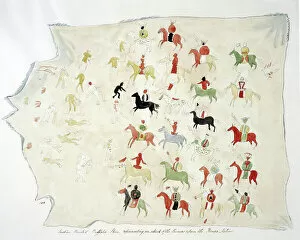
"Exploring the Intricacies: Unveiling Internal Anatomy through Art and Science" Delving into the hidden world of internal anatomy, we are captivated by the wonders that lie beneath our skin. From the delicate cross-section of a Honey Bee (Apis mellifera) to the meticulous illustrations of the Channel Tunnel Project, each piece unravels a unique perspective on this captivating subject. Intriguingly, even centuries ago, artists like Giovanni Battista Bianchi skillfully captured human form in terracotta sculptures, reminding us of our intricate inner workings. Meanwhile, Andreas Vesalius revolutionized anatomical understanding with his groundbreaking studies during the Renaissance era. As we journey through time and mediums, Waldeck's original artwork takes us back to Mesoamerica in 1831. The prints from "La nature et l'homme" by Rengade depict twin fetuses in their ideal position during gemellar pregnancy – a testament to both artistry and scientific observation. The diversity within these artworks is astounding; from portraits such as Lucrezia Panciatichi's enigmatic gaze immortalized in oil on wood panel to Giulia di Alessandro de Medici holding a book – perhaps symbolizing knowledge about one's own body. Even photography joins this exploration as two male nudes perch upon a rock in an evocative black-and-white image from 1890. This photograph captures not only physicality but also invites contemplation on vulnerability and strength intertwined within our internal structures. From ancient times to modern endeavors like the Channel Tunnel Project illustration from 1906, humanity has sought to unravel its own mysteries. Through art and science alike, we continue to marvel at our internal anatomy – an ever-evolving tapestry waiting for discovery.