
Internal Organ Collection

"Exploring the Intricacies Within

Background imageInternal Organ Collection: Metropolitan Cathedral Pipe Organs

Metropolitan Cathedral Pipe Organs
Metropolitan Cathedral Pipe Organ, view from bellow

Background imageInternal Organ Collection: Pig anatomy, artwork

Pig anatomy, artwork
Pig anatomy. Artwork showing the internal and external anatomy of the domesticated pig (Sus scrofa domestica), which is reared on farms worldwide for its meat

Background imageInternal Organ Collection: Human heart, artwork

Human heart, artwork
Human heart, computer artwork

Background imageInternal Organ Collection: Healthy heart, artwork F006 / 2957

Healthy heart, artwork F006 / 2957
Healthy heart. Computer artwork showing the right atrium of the heart

Background imageInternal Organ Collection: Breast, tongue, skin and finger sections. Lithograph of Corbie, based on a drawing by Leveille

Breast, tongue, skin and finger sections. Lithograph of Corbie, based on a drawing by Leveille
FLO4672795 Breast, tongue, skin and finger sections. Lithograph of Corbie, based on a drawing by Leveille, in Petity Atlas complet d'Anatomie descriptive du Corps Humain, by Dr

Background imageInternal Organ Collection: Neck, tongue and throat muscles. Lithograph of Choubard, based on a drawing by Leveille

Neck, tongue and throat muscles. Lithograph of Choubard, based on a drawing by Leveille
FLO4672612 Neck, tongue and throat muscles. Lithograph of Choubard, based on a drawing by Leveille, in Petity Atlas complet d'Anatomie descriptive du Corps Humain, by Dr

Background imageInternal Organ Collection: Frontispice of ' De succo pancreatico', Leiden, 1671, by Regnier (Regnerus) de Graaf (1641 1673)

Frontispice of " De succo pancreatico", Leiden, 1671, by Regnier (Regnerus) de Graaf (1641 1673)
XEE5029587 Frontispice of " De succo pancreatico", Leiden, 1671, by Regnier (Regnerus) de Graaf (1641 1673) Dutch doctor - Engraving of 1672; (add.info)

Background imageInternal Organ Collection: Frontispice de' De lactibus, sive, Lacteis venis, quarto vasorum mesaraicorum genere

Frontispice de" De lactibus, sive, Lacteis venis, quarto vasorum mesaraicorum genere, novo invento" de 1628
XEE5027828 Frontispice de" De lactibus, sive, Lacteis venis, quarto vasorum mesaraicorum genere, novo invento" de 1628

Background imageInternal Organ Collection: Internal muscles of the back, the deepest. Lithograph by Davesne, based on a drawing by Leveille

Internal muscles of the back, the deepest. Lithograph by Davesne, based on a drawing by Leveille
FLO4672659 Internal muscles of the back, the deepest. Lithograph by Davesne, based on a drawing by Leveille, in Petity Atlas complet d'Anatomie descriptive du Corps Humain, by Dr

Background imageInternal Organ Collection: Frontal bone of the skull. Lithograph of Corbie, in Petity Atlas complet d'Anatomie descriptive du

Frontal bone of the skull. Lithograph of Corbie, in Petity Atlas complet d'Anatomie descriptive du Corps Humain
FLO4672402 Frontal bone of the skull. Lithograph of Corbie, in Petity Atlas complet d'Anatomie descriptive du Corps Humain, by Dr

Background imageInternal Organ Collection: Muscles of the face, eyes and neck. Lithograph by Davesne, based on a drawing by Leveille

Muscles of the face, eyes and neck. Lithograph by Davesne, based on a drawing by Leveille
FLO4672617 Muscles of the face, eyes and neck. Lithograph by Davesne, based on a drawing by Leveille, in Petity Atlas complet d'Anatomie descriptive du Corps Humain, by Dr

Background imageInternal Organ Collection: Olfactory and maxillary nerves. Lithograph by Davesne, based on a drawing by Leveille

Olfactory and maxillary nerves. Lithograph by Davesne, based on a drawing by Leveille
FLO4672503 Olfactory and maxillary nerves. Lithograph by Davesne, based on a drawing by Leveille, in Petity Atlas complet d'Anatomie descriptive du Corps Humain, by Dr

Background imageInternal Organ Collection: Saphene vein and medial plantar nerves. Lithograph of Corbie, based on a drawing by Leveille

Saphene vein and medial plantar nerves. Lithograph of Corbie, based on a drawing by Leveille
FLO4672470 Saphene vein and medial plantar nerves. Lithograph of Corbie, based on a drawing by Leveille, in Petity Atlas complet d'Anatomie descriptive du Corps Humain, by Dr

Background imageInternal Organ Collection: Microscopic views of human spermatozoa. View of the animalculae or organic particles in the semen

Microscopic views of human spermatozoa. View of the animalculae or organic particles in the semen
FLO4714918 Microscopic views of human spermatozoa. View of the animalculae or organic particles in the semen. Handcoloured copperplate engraving by J

Background imageInternal Organ Collection: Hip muscles - 1866 illustration showing the muscles (brown), tendons (white) and bones (beige)

Hip muscles - 1866 illustration showing the muscles (brown), tendons (white) and bones (beige) of the human hip (centre)
JAB4119507 Hip muscles - 1866 illustration showing the muscles (brown), tendons (white) and bones (beige) of the human hip (centre) - Seen here are: Fig 1: sacrum and coccyx (tail bone); Fig 2

Background imageInternal Organ Collection: Chest and neck muscles - 1866 anatomical illustration showing the structure of the muscles (brown)

Chest and neck muscles - 1866 anatomical illustration showing the structure of the muscles (brown) and tendons (white)
JAB4119483 Chest and neck muscles - 1866 anatomical illustration showing the structure of the muscles (brown) and tendons (white) found in the rear of the human head (top) and neck (upper centre)

Background imageInternal Organ Collection: Pelvic and abdominal muscles - 1866 illustration of the internal muscles (brown) and tendons (white)

Pelvic and abdominal muscles - 1866 illustration of the internal muscles (brown) and tendons (white)
JAB4119447 Pelvic and abdominal muscles - 1866 illustration of the internal muscles (brown) and tendons (white) around the human pelvis (lower centre) and abdomen

Background imageInternal Organ Collection: Rib cage anatomy - 1866 illustration of the muscles (brown), tendons (white) and bones (beige)

Rib cage anatomy - 1866 illustration of the muscles (brown), tendons (white) and bones (beige) of the human rib cage
JAB4119446 Rib cage anatomy - 1866 illustration of the muscles (brown), tendons (white) and bones (beige) of the human rib cage, showing the sternum (breast bone, Fig 1), ribs

Background imageInternal Organ Collection: Leg and knee muscles - 1866 anatomical illustration showing the muscles (brown) and tendons (white)

Leg and knee muscles - 1866 anatomical illustration showing the muscles (brown) and tendons (white)
JAB4119438 Leg and knee muscles - 1866 anatomical illustration showing the muscles (brown) and tendons (white) of the human leg around the knee (centre) - Outer area of the knee - Vast inner muscle

Background imageInternal Organ Collection: Diaphragm anatomy - 1866 anatomical illustration showing the human diaphragm (top: centre)

Diaphragm anatomy - 1866 anatomical illustration showing the human diaphragm (top: centre)
JAB4119430 Diaphragm anatomy - 1866 anatomical illustration showing the human diaphragm (top: centre, bottom: upper-centre)

Background imageInternal Organ Collection: Arm muscles - 1866 illustration showing the structure of the muscles (brown) and tendons (white)

Arm muscles - 1866 illustration showing the structure of the muscles (brown) and tendons (white)
JAB4119402 Arm muscles - 1866 illustration showing the structure of the muscles (brown) and tendons (white) of the human arm - The internal

Background imageInternal Organ Collection: Pelvis anatomy - 1866 illustration showing the anatomy of the male (top) and female (bottom)

Pelvis anatomy - 1866 illustration showing the anatomy of the male (top) and female (bottom)
JAB4119375 Pelvis anatomy - 1866 illustration showing the anatomy of the male (top) and female (bottom) pelvis - Aponevroses abdominales, posterior plane - Fig 1: Crete iliaque Fig 2: Crete iliaque

Background imageInternal Organ Collection: Human hand - 1866 anatomical illustration showing the muscles (brown) and tendons (white)

Human hand - 1866 anatomical illustration showing the muscles (brown) and tendons (white) of the upper (right)
JAB4119374 Human hand - 1866 anatomical illustration showing the muscles (brown) and tendons (white) of the upper (right) and lower (left)

Background imageInternal Organ Collection: Lung anatomy 1866 illustrations showing the external anatomy of the human lungs (beige)

Lung anatomy 1866 illustrations showing the external anatomy of the human lungs (beige)
JAB4119246 Lung anatomy 1866 illustrations showing the external anatomy of the human lungs (beige), with the bronchi and trachea (white)

Background imageInternal Organ Collection: Male perineum 1866 illustration showing the muscles and other structures of the male perineum

Male perineum 1866 illustration showing the muscles and other structures of the male perineum - the region of the body
JAB4119197 Male perineum 1866 illustration showing the muscles and other structures of the male perineum - the region of the body located between the scrotum and anus Le perinee de l'homme

Background imageInternal Organ Collection: Prostate gland 1866 illustrations showing the structure of the prostate gland Prostate structure

Prostate gland 1866 illustrations showing the structure of the prostate gland Prostate structure - Fig 1
JAB4119151 Prostate gland 1866 illustrations showing the structure of the prostate gland Prostate structure - Fig 1: Prostate length cut Fig 2: Middle section cut

Background imageInternal Organ Collection: Brain sections, cerebellum and ventricles. Lithograph by Davesne, based on a drawing by Leveille

Brain sections, cerebellum and ventricles. Lithograph by Davesne, based on a drawing by Leveille
FLO4673145 Brain sections, cerebellum and ventricles. Lithograph by Davesne, based on a drawing by Leveille, in Petity Atlas complet d'Anatomie descriptive du Corps Humain, by Dr

Background imageInternal Organ Collection: Cross sections of the brain. Lithograph of Corbie, based on a drawing by Leveille

Cross sections of the brain. Lithograph of Corbie, based on a drawing by Leveille
FLO4673108 Cross sections of the brain. Lithograph of Corbie, based on a drawing by Leveille, in Petity Atlas complet d'Anatomie descriptive du Corps Humain, by Dr

Background imageInternal Organ Collection: Lymphatic system of the abdomen. Lithograph of Corbie, based on a drawing by Leveille

Lymphatic system of the abdomen. Lithograph of Corbie, based on a drawing by Leveille
FLO4673043 Lymphatic system of the abdomen. Lithograph of Corbie, based on a drawing by Leveille, in Petity Atlas complet d'Anatomie descriptive du Corps Humain, by Dr

Background imageInternal Organ Collection: Pelvis and arm circulatory system. Lithograph by Davesne, based on a drawing by Leveille

Pelvis and arm circulatory system. Lithograph by Davesne, based on a drawing by Leveille
FLO4672946 Pelvis and arm circulatory system. Lithograph by Davesne, based on a drawing by Leveille, in Petity Atlas complet d'Anatomie descriptive du Corps Humain, by Dr

Background imageInternal Organ Collection: Circulatory system of the abdomen. Lithograph of Corbie, based on a drawing by Leveille

Circulatory system of the abdomen. Lithograph of Corbie, based on a drawing by Leveille
FLO4672925 Circulatory system of the abdomen. Lithograph of Corbie, based on a drawing by Leveille, in Petity Atlas complet d'Anatomie descriptive du Corps Humain, by Dr

Background imageInternal Organ Collection: The circulatory system of the brain. Lithograph of Choubard, based on a drawing by Leveille

The circulatory system of the brain. Lithograph of Choubard, based on a drawing by Leveille
FLO4672884 The circulatory system of the brain. Lithograph of Choubard, based on a drawing by Leveille, in Petity Atlas complet d'Anatomie descriptive du Corps Humain, by Dr

Background imageInternal Organ Collection: Sections of the heart. Lithograph of Corbie, based on a drawing by Leveille

Sections of the heart. Lithograph of Corbie, based on a drawing by Leveille
FLO4672832 Sections of the heart. Lithograph of Corbie, based on a drawing by Leveille, in Petity Atlas complet d'Anatomie descriptive du Corps Humain, by Dr

Background imageInternal Organ Collection: Varietes of skin disease autoimmune psoriasis - Lithography by John Pass, published in 'Encyclopedia

Varietes of skin disease autoimmune psoriasis - Lithography by John Pass, published in "Encyclopedia
FLO4618788 Varietes of skin disease autoimmune psoriasis - Lithography by John Pass, published in "Encyclopedia Londinensis" by John Wilkes, edition J.Adlard, London

Background imageInternal Organ Collection: Scalp diseases, porrigo varietes and skin diseases: ulcerous pyodermatitis caused by bacteria

Scalp diseases, porrigo varietes and skin diseases: ulcerous pyodermatitis caused by bacteria - Lithography by John
FLO4618780 Scalp diseases, porrigo varietes and skin diseases: ulcerous pyodermatitis caused by bacteria - Lithography by John Pass, published in "Encyclopedia Londinensis" by John Wilkes

Background imageInternal Organ Collection: Adult crane desarticule 1867

Adult crane desarticule 1867
JAB4120023 Adult crane desarticule 1867; (add.info.: Adult crane desarticule 1867); Photo ©Jaime Abecasis

Background imageInternal Organ Collection: Bladder anatomy 1866 illustrations showing the internal (top) and external (bottom)

Bladder anatomy 1866 illustrations showing the internal (top) and external (bottom)
JAB4119242 Bladder anatomy 1866 illustrations showing the internal (top) and external (bottom) anatomy of the male urinary bladder, showing its muscular coating (brown)

Background imageInternal Organ Collection: Voice box and thyroid 1866 illustration showing the anatomy of the larynx (voice box)

Voice box and thyroid 1866 illustration showing the anatomy of the larynx (voice box) and thyroid (brown)
JAB4119213 Voice box and thyroid 1866 illustration showing the anatomy of the larynx (voice box) and thyroid (brown, lower centre) Shown here are the hyoid bone (A), thyroid cartilage (B)

Background imageInternal Organ Collection: Chest muscles - 1866 illustration showing the structure of the muscles (brown) and tendons (white)

Chest muscles - 1866 illustration showing the structure of the muscles (brown) and tendons (white) of the human chest
JAB4119479 Chest muscles - 1866 illustration showing the structure of the muscles (brown) and tendons (white) of the human chest

Background imageInternal Organ Collection: Leg and foot muscles - 1866 anatomical illustration showing the muscles (brown) and tendons (white)

Leg and foot muscles - 1866 anatomical illustration showing the muscles (brown) and tendons (white)
JAB4119476 Leg and foot muscles - 1866 anatomical illustration showing the muscles (brown) and tendons (white) of the right foot (left)

Background imageInternal Organ Collection: Shoulder muscles - 1866 illustration of the muscles (brown) and tendons (white)

Shoulder muscles - 1866 illustration of the muscles (brown) and tendons (white)
JAB4119462 Shoulder muscles - 1866 illustration of the muscles (brown) and tendons (white) associated with the human collarbone (clavicle, top) and shoulder blade (scapula)

Background imageInternal Organ Collection: Chest and shoulder muscles - 1866 anatomical illustration showing the structure of the muscles

Chest and shoulder muscles - 1866 anatomical illustration showing the structure of the muscles (brown)
JAB4119433 Chest and shoulder muscles - 1866 anatomical illustration showing the structure of the muscles (brown) and tendons (white)

Background imageInternal Organ Collection: Hand muscles - 1866 anatomical illustration showing the muscles (brown) and tendons (white)

Hand muscles - 1866 anatomical illustration showing the muscles (brown) and tendons (white) of the human hand
JAB4119426 Hand muscles - 1866 anatomical illustration showing the muscles (brown) and tendons (white) of the human hand, with insets (left)

Background imageInternal Organ Collection: Rib cage anatomy - 1866 illustration showing the bones of the human rib cage

Rib cage anatomy - 1866 illustration showing the bones of the human rib cage, including the ribs (bottom)
JAB4119411 Rib cage anatomy - 1866 illustration showing the bones of the human rib cage, including the ribs (bottom), spine (centre-left) and sternum (breast bone, centre and upper right) Le Thorax

Background imageInternal Organ Collection: Pelvis anatomy - 1866 illustration showing the anatomy of the male (top) and female (bottom)

Pelvis anatomy - 1866 illustration showing the anatomy of the male (top) and female (bottom) pelvis - The pelvis
JAB4119404 Pelvis anatomy - 1866 illustration showing the anatomy of the male (top) and female (bottom) pelvis - The pelvis, anatomy, osteology - Fig 1: female pelvis Fig 2: male pelvis, Musculotion

Background imageInternal Organ Collection: Hip muscles - 1866 illustration showing the structure of the muscles (brown) and tendons (white)

Hip muscles - 1866 illustration showing the structure of the muscles (brown) and tendons (white)
JAB4119401 Hip muscles - 1866 illustration showing the structure of the muscles (brown) and tendons (white) of the upper right leg and hip

Background imageInternal Organ Collection: Arm muscles - 1866 illustration showing the structure of the muscles (brown) and tendons (white)

Arm muscles - 1866 illustration showing the structure of the muscles (brown) and tendons (white)
JAB4119391 Arm muscles - 1866 illustration showing the structure of the muscles (brown) and tendons (white) of the human arm and top of the hand (bottom left and right)

Background imageInternal Organ Collection: Abdominal muscles - 1866 illustration showing the structure of the muscles (brown)

Abdominal muscles - 1866 illustration showing the structure of the muscles (brown) and tendons (white)
JAB4119387 Abdominal muscles - 1866 illustration showing the structure of the muscles (brown) and tendons (white) found in the rear of the human trunk - L'abdomen - Anatomy - Region anterieure de


For sale as Licensed Images

Choose your image, Select your licence and Download the media

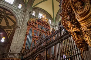
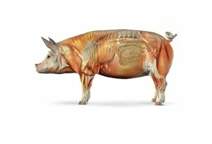
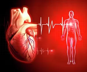
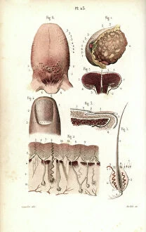
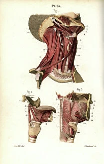
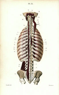
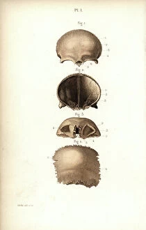
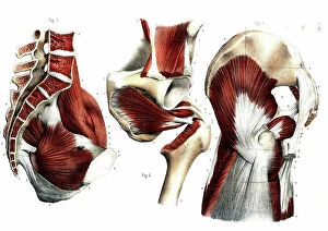
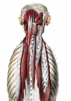
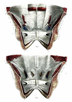
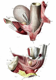
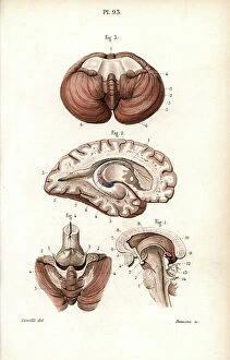
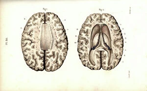
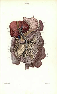
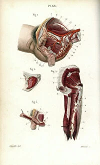
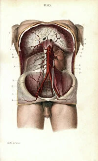
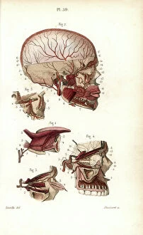
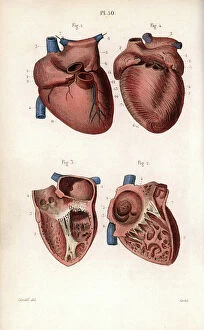
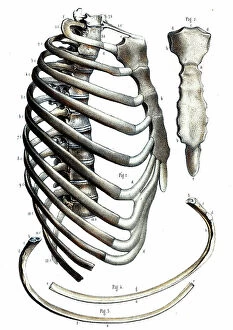
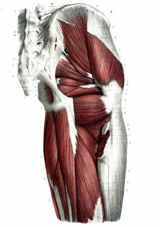
"Exploring the Intricacies Within: A Journey into Internal Organ Artwork" Delve into the fascinating world of internal organs through captivating artwork that unveils their hidden wonders. From pig anatomy to the majestic Metropolitan Cathedral Pipe Organs, this collection takes you on an artistic voyage like no other. Witness the intricate beauty of a human heart depicted in stunning artwork, reminding us of its vital role in sustaining life. Marvel at a healthy heart portrayed in exquisite detail, symbolizing the importance of maintaining cardiovascular well-being (F006 / 2957). Discover the complex network of coronary arteries illustrated with precision, offering insight into how these vessels nourish our hearts and keep them beating strong. Uncover the secrets held within a lung's X-ray image, revealing both its delicate structure and remarkable functionality. Behold the mesmerizing Mashpi glassfrog from Ecuador, showcasing its translucent underside where internal organs become visible. This captive creature provides a rare glimpse into nature's intricacy and diversity. Explore further as we unravel the mysteries of the human cardiovascular system—a masterpiece designed for efficient circulation and oxygenation throughout our bodies. Delight in an illustration depicting a baby's heart and circulatory system—reminding us that even from infancy, our hearts are at work to sustain life. Immerse yourself in breathtaking artwork capturing every aspect of our intricate cardiovascular system—an awe-inspiring testament to human resilience and vitality. Let your mind wander through vibrant depictions of our brain—the command center orchestrating all bodily functions with unparalleled complexity. Transport yourself back in time with a vintage depiction showing a median section of a cow—revealing principal organs responsible for digestion and more (c1905). Witness how even animals possess inner workings worth exploring. Embark on this visual journey celebrating internal organ artistry—a harmonious blend between science and creativity that invites us to appreciate both their aesthetic appeal and indispensable roles within ourselves.