
Intestinal Villi Collection
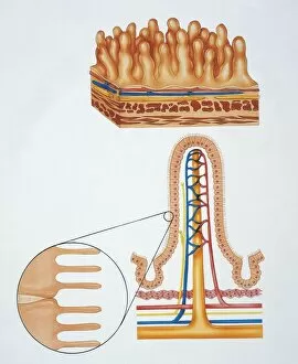
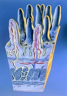
Intestinal villi, the tiny finger-like projections lining the walls of our small intestine, play a crucial role in nutrient absorption


For sale as Licensed Images
Choose your image, Select your licence and Download the media
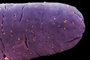
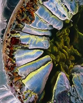
Intestinal villi, the tiny finger-like projections lining the walls of our small intestine, play a crucial role in nutrient absorption. These intricate structures are like nature's own microscopic work of art. Through illustrations and artwork such as F007/7807 and F007/7808, we can visualize the complexity and beauty of intestinal villi. Their delicate yet efficient design allows for maximum surface area within our digestive system. In a light micrograph capturing the colon lining, we witness the intricacy of these villi up close. The image showcases their unique shape and arrangement, providing insight into their function in absorbing water and electrolytes from digested food. Coloured SEM images further highlight the remarkable structure of these villi. From sectioned pieces in the ileum to those found in different parts of the small intestine like jejunum, each image reveals their vibrant colors that make them stand out under magnification. The false-colour SEM through the wall of duodenum gives us an extraordinary glimpse into how these villi extend into our intestines' inner layers. This visualization helps us understand how they facilitate nutrient absorption by increasing surface area contact with chyme (partially digested food). As we explore more SEM images showcasing individual villi from various angles, it becomes evident why they are essential for digestion. Their presence ensures efficient absorption by maximizing contact between nutrients and absorptive cells present on their surfaces.