
Intestines Collection (#2)
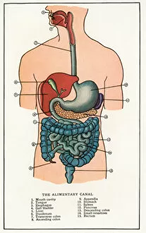
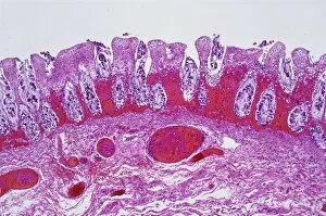
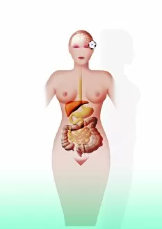
Intestines, the intricate network of tubes and tissues that play a vital role in various organisms' digestive systems


For sale as Licensed Images
Choose your image, Select your licence and Download the media
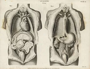
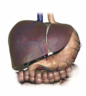
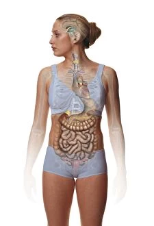
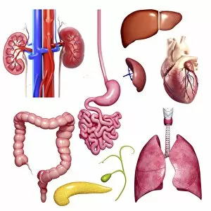
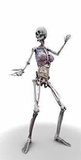
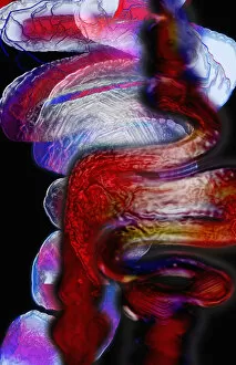
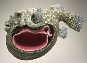
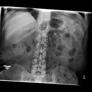
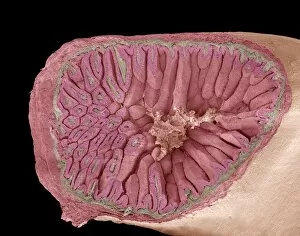
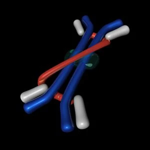
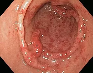
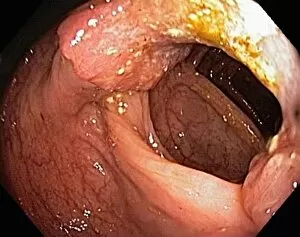
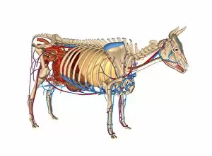
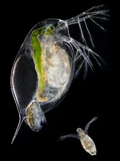
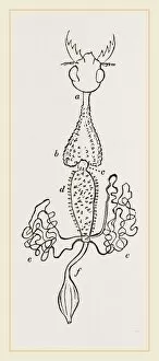
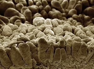
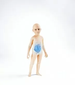
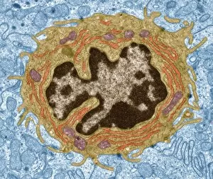
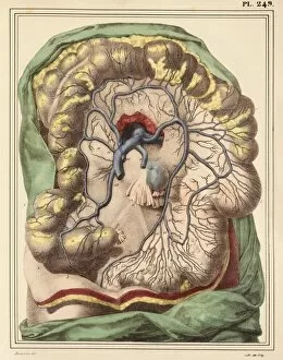
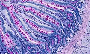
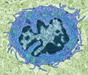
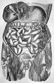
Intestines, the intricate network of tubes and tissues that play a vital role in various organisms' digestive systems. From bees to humans, these remarkable organs are essential for nutrient absorption and waste elimination. In the world of bee anatomy, the intestines serve as a crucial part of their digestive process. Through stunning artwork, we can explore the delicate intricacies of these tiny creatures' intestinal structure. Moving on to larger animals like pigs, their small intestine takes center stage in absorbing nutrients from food. Scanning electron microscopy (SEM) allows us to delve into its microscopic details and marvel at nature's precision. E. Coli bacteria may often be associated with illness, but through SEM imagery, we witness their presence within our own intestines. Understanding this relationship is key to maintaining a healthy human digestive system. Horses possess an impressive gastrointestinal tract too; exploring the anatomy of their intestines reveals how they efficiently break down plant matter for digestion. Artwork showcasing this complexity brings forth admiration for these magnificent creatures. When it comes to beef carcasses, examining the intestines provides valuable insights into meat quality and safety standards during processing stages. However, not all inhabitants within our they can beneficial; intestinal protozoan parasites pose health risks worldwide. Transmission electron microscopy (TEM) captures detailed images of Cryptosporidium protozoa responsible for causing infections in humans and animals alike. Zooming even closer under TEM reveals fascinating structures called intestinal microvilli lining our gut walls – finger-like projections that increase surface area for nutrient absorption. Shifting perspectives towards human internal organ anatomy from behind unveils how interconnected our they can with other vital systems such as kidneys or lungs – emphasizing their significance beyond digestion alone. Even dogs have unique anatomical features worth exploring. Artistic representations allow us to appreciate canine intestinal complexities while acknowledging similarities and differences compared to other species studied before.