Invertebrates Collection (#97)

"Invertebrates

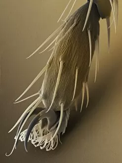
Background imageInvertebrates Collection: Vine weevil foot, SEM

Vine weevil foot, SEM
Weevil foot. Coloured scanning electron micrograph (SEM) of the foot of a black vine weevil (Otiorhynchus sulcatus). The foot comprises a pair of adhesive hairy pads (lower centre)

Background imageInvertebrates Collection: Butterfly eggs

Butterfly eggs. Coloured scanning electron micrograph (SEM) of eggs of the large white (cabbage white) butterfly Pieris brassicae

Background imageInvertebrates Collection: Brassica weevil, SEM

Brassica weevil, SEM
Brassica weevil (Ceutorhynchus sp.), coloured scanning electron micrograph (SEM). This beetle, which grows up to 3 millimetres in length, is a major pest of cabbages (Brassica sp.)

Background imageInvertebrates Collection: Mussel glue threads, SEM

Mussel glue threads, SEM
Mussel glue. Coloured scanning electron micrograph (SEM) of the byssus (glue threads) of a common mussel (Mytilus edulis)
Background imageInvertebrates Collection: Glasswing butterflies

Glasswing butterflies (Greta oto). This semi- transparent butterfly is native to Central America. It is not easy to see because of its partially transparent wings

Background imageInvertebrates Collection: Julia butterfly (Dryas julia)

Julia butterfly (Dryas julia). This butterfly can be found in South and Central America

Background imageInvertebrates Collection: Lacewing butterfly

Lacewing butterfly (Cethosia cyane). The bright colouration of this butterfly is a warning to prospective predators that it is unpalatable

Background imageInvertebrates Collection: Red wood ant

Red wood ant. Coloured scanning electron micrograph (SEM) of a red wood ant (Formica rufa). Its antennae are seen at upper left and right. Behind these on its head are its eyes

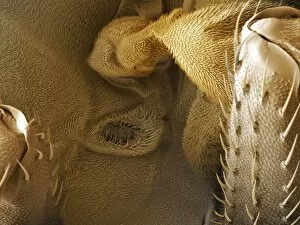
Background imageInvertebrates Collection: Female bedbugs abdomen, SEM

Female bedbugs abdomen, SEM
Female bedbugs abdomen. Scanning electron micrograph (SEM) of the external groove over the pleural membrane on the abdomen of a female bedbug (Cimex lectularius)

Background imageInvertebrates Collection: Green stink bug

Green stink bug (Nezara viridula) on a flower. This insect is a pest of cereal crops. Stink bugs derive their name from the foul-smelling secretion which they emit from their thoraic glands as a

Background imageInvertebrates Collection: Mealworm beetles mating

Mealworm beetles mating. Coloured scanning electron micrograph (SEM) of yellow mealworm beetles (Tenebrio molitor) mating

Background imageInvertebrates Collection: Wasp

Wasp (Family Vespidae). This is one over 4, 000 species of social wasp. Social wasps are distinguished from solitary wasps by the resting position of their wings

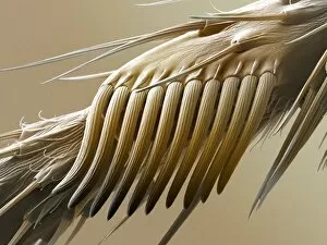
Background imageInvertebrates Collection: Fruit fly sex comb, SEM

Fruit fly sex comb, SEM
Fruit fly (Drosophila melanogaster) sex comb, coloured scanning electron micrograph (SEM). The sex comb is found only on the front legs of males

Background imageInvertebrates Collection: Small flambeau butterfly

Small flambeau butterfly (Eueides heliconius). This butterfly feeds on passion flower plants (Passiflora sp.). it is found in the tropics of South America and Trinidad

Background imageInvertebrates Collection: Hen flea, SEM

Hen flea, SEM
Hen flea (Ceratophyllus gallinae), coloured scanning electron micrograph. This parasitic insect inhabits the nests of birds, usually those that nest in holes

Background imageInvertebrates Collection: Glasswing butterfly

Glasswing butterfly (Greta oto) feeding from Lantana flowers (Lantana camera). This semi- transparent butterfly is native to Central America

Background imageInvertebrates Collection: Gall mites, SEM

Gall mites, SEM
Gall mites. Coloured scanning electron micrograph (SEM) of several gall mites (Eriophyes sp.). These pests infest a wide range of plants, causing economic damage to food crops and forests

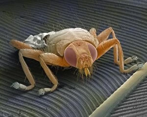
Background imageInvertebrates Collection: Parasitic fly, SEM Z340 / 0605

Parasitic fly, SEM Z340 / 0605
Parasitic fly. Coloured scanning electron micrograph (SEM) of the fly Ornithomyia avicularia. This is a parasite of nesting birds, especially house martins and swallows

Background imageInvertebrates Collection: Fruit fly antenna, SEM

Fruit fly antenna, SEM
Fruit fly antenna. Coloured scanning electron micrograph (SEM) of the head of a male fruit fly (Drosophila melanogaster). Seen here is an antenna (left) and part of a compound eye (upper right, red)

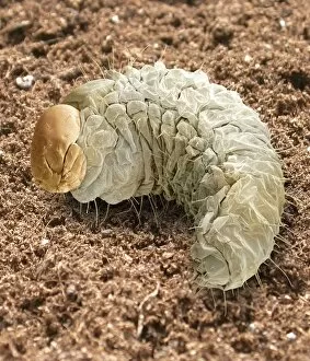
Background imageInvertebrates Collection: Vine weevil larva, SEM

Vine weevil larva, SEM
Black vine weevil larva (Otiorhynchus sulcatus), coloured scanning electron micrograph (SEM). This insect is a pest that attacks a wide range of plants, including the yew, rhododendron, azalea

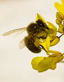
Background imageInvertebrates Collection: Honey bee feeding

Honey bee feeding
Honey bee (Apis mellifera) feeding on a flower. This is a field bee, an older female worker bee who spends time out of the colony foraging for food

Background imageInvertebrates Collection: Coloured SEM of a shield bug (Elasmostethus sp. )

Coloured SEM of a shield bug (Elasmostethus sp. )
Shield bug feeding. Coloured scanning electron micrograph (SEM) of a shield bug (Elasmostethus interstinctus) feeding on a leaf

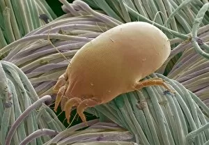
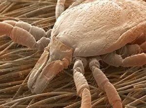
Background imageInvertebrates Collection: Dust mite

Dust mite. Coloured scanning electron micrograph (SEM) of the house dust mite, Dermatophagoides pteronyssinus on woven household fabric. This mite has eight legs, with the head at lower left

Background imageInvertebrates Collection: Coloured SEM of hamuli of honey bee wing

Coloured SEM of hamuli of honey bee wing
Honeybee wing coupling mechanism. Coloured Scanning Electron Micrograph of the edge of the wing of a Honey Bee, Apis mellifera

Background imageInvertebrates Collection: Snail, SEM

Snail, SEM
Snail. Coloured scanning electron micrograph (SEM) of a laver spire snail (Hydrobia ulvae). The snail can seal itself inside its shell by pulling a plug (brown) over its operculum (lower right)

Background imageInvertebrates Collection: Centipede, SEM

Centipede, SEM
Garden centipede. Coloured scanning electron micrograph (SEM) of the head and upper body of a garden centipede (Lithobius sp.)

Background imageInvertebrates Collection: Coloured SEM of Rose aphid nymph feeding on a leaf

Coloured SEM of Rose aphid nymph feeding on a leaf
Rose aphid nymph. Coloured scanning electron micrograph (SEM) of a Rose aphid nymph Macrosiphon rosae feeding on a rose leaf. This wingless nymph is an immature form of the adult aphid

Background imageInvertebrates Collection: Coloured SEM of a crane fly (Tipula maxima)

Coloured SEM of a crane fly (Tipula maxima)
Crane fly. Coloured scanning electron micrograph (SEM) of a female crane fly (Tipula maxima), or daddy long legs. Its head is at centre left

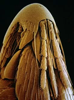
Background imageInvertebrates Collection: Bird flea head, SEM

Bird flea head, SEM
Head of a bird flea. False-colour scanning electron micrograph (SEM) of the head of the bird flea, Ceratophylus galinae. The flea is a parasite on dry nesting birds, such as the Blue Tit

Background imageInvertebrates Collection: Squid suckers, SEM

Squid suckers, SEM
Squid suckers. Coloured scanning electron micrograph (SEM) of suckers on the tentacles of a common squid (Loligo forbesi). The suckers are cup-like structures that are used to grasp prey

Background imageInvertebrates Collection: Fruit fly head, SEM

Fruit fly head, SEM
Fruit fly head. Coloured scanning electron micrograph (SEM) of the head of a male fruit fly (Drosophila melanogaster). Its two compound eyes (red) are seen on either side of the head

Background imageInvertebrates Collection: Deer tick, SEM

Deer tick, SEM
Deer tick. Coloured scanning electron micrograph (SEM) of the head of a deer tick (Ixodes ricinus), a bloodsucking parasite of animals and humans

Background imageInvertebrates Collection: Fruit fly foot, SEM

Fruit fly foot, SEM
Fruit fly foot. Coloured scanning electron micrograph (SEM) of the foot of a male fruit fly (Drosophila melanogaster). The two claws are used to grip rough surfaces

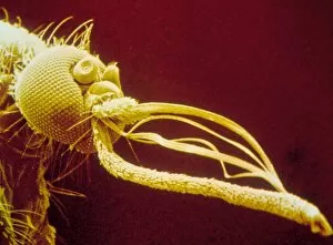
Background imageInvertebrates Collection: Coloured SEM of head of Aedes punctor mosquito

Coloured SEM of head of Aedes punctor mosquito
Head of a mosquito. Coloured scanning electron micrograph of the head of a female yellow fever mosquito, Aedes punctor. Like other insects, the mosquito has compound eyes, consisting of many facets

Background imageInvertebrates Collection: Ant communication, SEM

Ant communication, SEM
Ant communication. Coloured scanning electron micrograph (SEM) of three wood or heathland ants (Formica fusca) communicating. Ants are social insects that form large colonies

Background imageInvertebrates Collection: Garden ground beetle, SEM

Garden ground beetle, SEM
Garden ground beetle. Coloured scanning electron micrograph (SEM) of a garden ground beetle (Notiophilus aquaticus). This beetle has huge protruding eyes (dark brown, centre)

Background imageInvertebrates Collection: Fruit fly spiracle, SEM

Fruit fly spiracle, SEM
Fruit fly (Drosophila melanogaster) spiracle. Coloured scanning electron micrograph (SEM) of the spiracle (air pore) of a male fruit fly

Background imageInvertebrates Collection: Dust mites

Dust mites. Coloured scanning electron micrograph (SEM) of two dust mites (Dermatophagoides sp.) on fabric fibres. Millions of dust mites inhabit the home, feeding on shed skin cells

Background imageInvertebrates Collection: Col. SEM of head of zebra jumping spider, Salticus

Col. SEM of head of zebra jumping spider, Salticus
Head of jumping spider. Coloured Scanning Electron Micrograph (SEM) of the head of a Zebra jumping spider, Salticus scenicus

Background imageInvertebrates Collection: Spider irritant hair

Spider irritant hair
Tarantula spider hairs. Coloured scanning electron micrograph (SEM) of the irritant hairs of a Mexican red-knee tarantula (Brachypelma smithi)

Background imageInvertebrates Collection: Sheep tick, SEM

Sheep tick, SEM
Sheep tick. Coloured scanning electron micrograph (SEM) of a sheep tick (Ixodes ricinus). This is a bloodsucking parasite of animals and humans

Background imageInvertebrates Collection: LM of Demodex folliculorum

LM of Demodex folliculorum
Light micrograph in interference contrast illumination of the Follicle Mite (Demodex folliculorum). Follicle Mites are human ectoparasites, aggregating around hair follicles and in the orifices

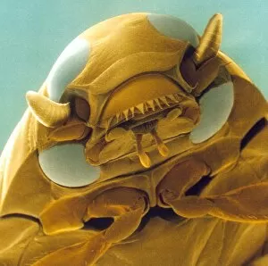
Background imageInvertebrates Collection: Head of a whirligig beetle, SEM

Head of a whirligig beetle, SEM
Whirligig beetle. Coloured scanning electron micrograph of the head of a whirligig beetle (Gyrinus natator). Whirligig beetles are small

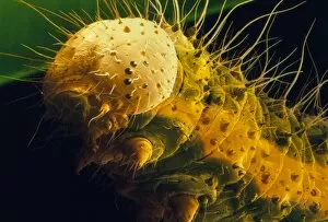
Background imageInvertebrates Collection: Cabbage white caterpillar, SEM

Cabbage white caterpillar, SEM
Cabbage white caterpillar. Coloured scanning electron micrograph (SEM) of the caterpillar (larva) of a large (or cabbage) white butterfly (Pieris brassicae)

Background imageInvertebrates Collection: Garden centipede, SEM

Garden centipede, SEM
Garden centipede. Coloured scanning electron micrograph (SEM) of a garden centipede (Lithobius sp.). A centipedes body is divided into segments, each containing a pair of legs

Background imageInvertebrates Collection: Great peacock moth

Great peacock moth (Saturnia pyri). This is the largest moth native to Europe. It has a wingspan of up to 15 centimetres and very distinctive ringed eye spots on its wings

Background imageInvertebrates Collection: Cowpea weevil

Cowpea weevil preparing for flight. Coloured scanning electron micrograph (SEM) of the cowpea weevil (Callosobruchus maculatus), also known as the seed beetle

Background imageInvertebrates Collection: Tiger longwing butterfly

Tiger longwing butterfly (Heliconius hecale). Colouring within this species of butterfly is highly variable, and cross-species mimicry is common


For sale as Licensed Images

Choose your image, Select your licence and Download the media

"Invertebrates: A Fascinating World of Diversity and Adaptation" From the intricate honeycombs built by diligent honey bees to the captivating sight of a lobster's inner workings revealed through an X-ray, invertebrates never cease to amaze us. The Red Admiral butterfly gracefully displays its vibrant wings, showcasing nature's artistic brilliance. Meanwhile, under the lens of a scanning electron microscope, calcareous phytoplankton reveal their delicate beauty as they float effortlessly in the vast ocean. In Kagoshima Prefecture, Kyushu, Japan, a male Leach's sea star steals the spotlight as it engages in broadcast spawning - an awe-inspiring phenomenon where streams of sperm are released from its arms into the water. This mesmerizing moment captured by a talented wildlife photographer earned them recognition as the winner in the Underwater category at Wildlife Photographer of the Year 2022. Venturing into Croatian waters along Korcula Island in the Adriatic Sea reveals another enchanting creature - a Nudibranch known as Janolus cristatus. Its vibrant colors and graceful movements make it a true gem beneath the waves. A sea green swallowtail butterfly flutters elegantly across meadows and gardens while fruit flies unveil their intricate structures when observed under high-resolution SEM imaging techniques. Delving deep into Earth's history brings us face-to-face with ancient wonders like trilobite fossils that offer glimpses into prehistoric times. These extinct arthropods remind us of our planet's rich evolutionary past. Witnessing an ocellate octopus swimming up from the depths is like witnessing magic unfold before our eyes. Its ethereal appearance leaves us captivated by nature's ability to create such extraordinary creatures. Lastly, we encounter a painted lady butterfly whose delicate patterns adorn fields with splashes of color and gracefulness. And even within microscopic realms lies astonishing complexity; for instance, observing dog tapeworm heads through SEM reveals the intricate structures that allow these parasites to thrive.