
Jaws Collection (#9)

"Jaws

Background imageJaws Collection: Trap-jaw ant carrying eggs C013 / 7056

Trap-jaw ant carrying eggs C013 / 7056
Trap-jaw ant carrying eggs. Close-up of a trap-jaw ant (Odontomachus bauri) using its large mandibles to carry eggs. This Central

Background imageJaws Collection: Trap-jaw ant carrying eggs C013 / 7055

Trap-jaw ant carrying eggs C013 / 7055
Trap-jaw ant carrying eggs. Close-up of a trap-jaw ant (Odontomachus bauri) using its large mandibles to carry eggs. This Central

Background imageJaws Collection: Trap-jaw ant C013 / 7054

Trap-jaw ant C013 / 7054
Trap-jaw ant. Close-up of a trap-jaw ant (Odontomachus bauri), showing its large mandibles (right) protruding to the left and right of the head

Background imageJaws Collection: Head and neck anatomy, artwork

Head and neck anatomy, artwork
Head and neck anatomy. Computer artwork of a human head and neck with the overlying tissues removed to show the brain (pink) within a sectioned skull that includes the teeth (white) of the jaws

Background imageJaws Collection: Spider stalking prey, SEM

Spider stalking prey, SEM
Spider stalking prey. Coloured scanning electron micrograph (SEM) of a wolf spider (Euophrys sp.) stalking a fly. Magnification: x19 when printed at 10 centimetres wide

Background imageJaws Collection: Spider - male - close-up of head showing fangs

Spider - male - close-up of head showing fangs
SPH-2358 Spider - male - close-up of head showing fangs Segestria florentina Segestria florentina is an introduced species. Its found in the south of England and Wales and in the vicinity of ports

Background imageJaws Collection: Dromaeosaurus, Fuzzy Raptor

Dromaeosaurus, Fuzzy Raptor
An animatronic model of the fuzzy raptor or dromaeosaurus created by Kokoro of Japan for the Natural History Museum. The fuzzy raptor was a small dinosaur covered in branched feathers

Background imageJaws Collection: Harvester Ant (Messor barbara) major and minor workers, major worker with jaws open in defensive

Harvester Ant (Messor barbara) major and minor workers, major worker with jaws open in defensive posture, Chaine des Alpilles, Bouches-du-Rhone, Provence, France, June

Background imageJaws Collection: Samsons capital. 12th c. Samson breaking the

Samsons capital. 12th c. Samson breaking the jaws of a lion. Romanesque art. Relief on rock. SPAIN. Barcelona. Frederic Mares Museum. Proc: SPAIN. Villaherreros

Background imageJaws Collection: Xochipilli. Prince of Flowers

Xochipilli. Prince of Flowers
Human head going out from animals jaws. Late classical period. Clay. Maya art. Sculpture on rock. Proc: MEXICO

Background imageJaws Collection: Gray / Grey / Timber Wolf - male yawning - controlled conditions

Gray / Grey / Timber Wolf - male yawning - controlled conditions
CAN-4318 Gray / Grey / Timber Wolf - male yawning Canis lupus controlled conditions John Cancalosi Please note that prints are for personal display purposes only and may not be reproduced in anyway

Background imageJaws Collection: Reef Lizardfish - opening mouth - Indonesia

Reef Lizardfish - opening mouth - Indonesia
LEE-261 Reef Lizardfish - opening mouth Indonesia Synodus variegatus Lea Lee contact details: prints@ardea.com tel: +44 (0) 20 8318 1401

Background imageJaws Collection: Tyrannosaurus Rex - teeth

Tyrannosaurus Rex - teeth
PM-8881 Dinosaur - Teeth of Tyrannosaurus Rex Pat Morris contact details: prints@ardea.com tel: +44 (0) 20 8318 1401

Background imageJaws Collection: Crocodile Shooting Party - Sudan

Crocodile Shooting Party - Sudan
How many Sudanese marksmen does it take to shoot a crocodile.... Date: circa 1910s

Background imageJaws Collection: Cuban Crocodile (Crocodylus rhombifer) adult, with mouth open, close-up of head (captive)

Cuban Crocodile (Crocodylus rhombifer) adult, with mouth open, close-up of head (captive)

Background imageJaws Collection: Wood Ant (Formica sp. ) adult workers, fighting, with severed head still grasping with mandibles

Wood Ant (Formica sp. ) adult workers, fighting, with severed head still grasping with mandibles, near Minerve
Wood Ant (Formica sp.) adult workers, fighting, with severed head still grasping with mandibles, near Minerve, Herault, Languedoc-Roussillon, France, may

Background imageJaws Collection: Gharial (Gavialis gangeticus) adult female, close-up of head, in shallow water at edge of river

Gharial (Gavialis gangeticus) adult female, close-up of head, in shallow water at edge of river, Chambal River, Uttar Pradesh, India, january

Background imageJaws Collection: Nomad Bee (Nomada lathburiana) adult female, sleeping, with jaws locked onto stem, Leicestershire

Nomad Bee (Nomada lathburiana) adult female, sleeping, with jaws locked onto stem, Leicestershire, England, april

Background imageJaws Collection: Nomad Bee (Nomada sp. ) adult, sleeping with jaws locked onto flower petal, Leicestershire

Nomad Bee (Nomada sp. ) adult, sleeping with jaws locked onto flower petal, Leicestershire, England, may
Nomad Bee (Nomada sp.) adult, sleeping with jaws locked onto flower petal, Leicestershire, England, may

Background imageJaws Collection: Mayfly nymph head, SEM

Mayfly nymph head, SEM
Mayfly nymph head, scanning electron micrograph (SEM). Mayflies (order Ephemeroptera) are the oldest surviving winged insects

Background imageJaws Collection: Earwig head, SEM

Earwig head, SEM
Earwig. Coloured scanning electron micrograph (SEM) of the head of an unidentified earwig (order Dermaptera). The bases of the long, slender antennae (purple, upper frame)

Background imageJaws Collection: Red fox eating a chick

Red fox eating a chick
Red fox (Vulpes vulpes) eating a chick. Red foxes are found in North America, Eurasia and North Africa, and are the only native wild canines remaining in the UK and Ireland

Background imageJaws Collection: Gorilla skull, X-ray

Gorilla skull, X-ray
Gorilla skull. Coloured X-ray of a front view of the skull of a male gorilla (Gorilla gorilla). The gorilla is the largest living primate

Background imageJaws Collection: Ant mouth, SEM

Ant mouth, SEM
Ant mouthparts. Coloured scanning electron micrograph (SEM) of the mouthparts of an ant (family Formicidae). The powerful mandibles (jaws, centre and upper left) are overlapping

Background imageJaws Collection: Damselfly nymph skin

Damselfly nymph skin. Light micrograph of the head and thorax of a shed skin from a damselfly nymph (Coenagrion sp.). This is the ventral (underside)

Background imageJaws Collection: Soldier ant jaws, SEM

Soldier ant jaws, SEM
Soldier ant jaws. Coloured scanning electron micrograph (SEM) of the head of a leaf-cutter ant (Atta sexdens). This is a soldier ant

Background imageJaws Collection: Sumatran stag beetle

Sumatran stag beetle (Dorcus titanus). This beetle has large antler-like jaws (mandibles, upper centre). These jaws change size and shape during the beetles life

Background imageJaws Collection: Bee mouthparts, light micrograph

Bee mouthparts, light micrograph. The two darker brown upper appendages are the mandibles (jaws), which are used for handling objects and for communicating with other bees

Background imageJaws Collection: Red wood ant, SEM

Red wood ant, SEM
Red wood ant. Coloured scanning electron micrograph (SEM) of the head of a red ant (Formica rufa). Its long sensory antennae are seen protruding from its head at lower centre and lower left

Background imageJaws Collection: Devils coach horse beetle, SEM

Devils coach horse beetle, SEM
Devils coach horse. Coloured scanning electron micrograph (SEM) of the underside of a devils coach horse beetle (Staphylinus olens)

Background imageJaws Collection: Head of a parasitic wasp, SEM

Head of a parasitic wasp, SEM
Head of a parasitic wasp. Coloured scanning electron micrograph (SEM) of the head of a parasitic wasp (Aphelinus abdominalis). This tiny wasp is a parasite of potato and foxglove aphids

Background imageJaws Collection: Fungus weevil head, SEM

Fungus weevil head, SEM
Weevil head. Coloured scanning electron micrograph (SEM) of the head of an endangered fungus weevil (Notioxenus sp.). The mouthparts are at lower right

Background imageJaws Collection: Loss of teeth, X-ray

Loss of teeth, X-ray
Loss of teeth. Coloured pan-oral X-ray of the mouth of an elderly man showing his few remaining teeth. Some of the teeth (for instance at centre right) have fillings (black) in them

Background imageJaws Collection: Complete loss of teeth, X-ray

Complete loss of teeth, X-ray
Loss of teeth. Coloured X-ray (side view) of the head of a 49 year old woman showing complete loss of teeth in both upper and lower jaws

Background imageJaws Collection: Paranasal sinuses, X-ray

Paranasal sinuses, X-ray
Paranasal sinuses. Coloured X-ray of a sagittal section through a human skull. The skull has been sliced in half down the centre and the regions of the paranasal sinuses coloured

Background imageJaws Collection: Skull, CT scan

Skull, CT scan
Human skull, coloured computed tomography (CT) scan. At top is the rounded cranium which houses the brain. The orbits (eye sockets) and nasal cavity (white) are seen

Background imageJaws Collection: Shark jaws

Shark jaws, coloured X-ray

Background imageJaws Collection: Jaws of a shark

Jaws of a shark, coloured X-ray

Background imageJaws Collection: Helicoprion prehistoric shark

Helicoprion prehistoric shark
Helicoprion. Artwork of the prehistoric shark Helicoprion. This genus of shark existed from the late Carboniferous period (280 million years ago)

Background imageJaws Collection: Water spider, SEM

Water spider, SEM
Water spider. Coloured scanning electron micrograph (SEM) of the head of a water spider (Argyroneta aquatica), showing its large jaws (mandibles, centre right) and its eyes (upper right). A

Background imageJaws Collection: Antlion larva, SEM

Antlion larva, SEM
Antlion larva, coloured scanning electron micrograph (SEM). The larval form of this insect (family Myrmeleontide) lives at the bottom of a cone shaped hole

Background imageJaws Collection: Boa constrictor eating a mouse C008 / 3368

Boa constrictor eating a mouse C008 / 3368
Boa constrictor (Boa constrictor) eating a mouse. A snakes jaws are very flexible, which enables them to swallow their prey whole. Boa constrictors are a popular species in the pet trade

Background imageJaws Collection: Lesser stag beetle

Lesser stag beetle (Dorcus parallelipipedus) on a path. Photographed in Kingcombe, Dorset, UK, in June

Background imageJaws Collection: Animal teeth comparison, artwork

Animal teeth comparison, artwork
Animal teeth comparison. Computer artwork comparing the tooth and jaw structure of a herbivore (top), a carnivore (middle), and a human (omnivore, bottom)

Background imageJaws Collection: Jumping spider jaws, SEM

Jumping spider jaws, SEM
Jumping spider jaws. Coloured scanning electron micrograph (SEM) of the head of a jumping spider (family Salticidae), showing its large jaws (mandibles, centre)

Background imageJaws Collection: Broken chin, 3D CT scan

Broken chin, 3D CT scan
Broken chin. Three-quarter side view 3D computed tomography (CT) scan of a patient with a broken chin. The lower jaw is fractured just below the bottom front incisor teeth

Background imageJaws Collection: Teeth, 3D CT scan

Teeth, 3D CT scan. Frontal view of a persons jaw and teeth with braces on the upper teeth. The wisdom teeth (upper left and upper right) have not yet penetrated the gums

Background imageJaws Collection: Brain and skull, artwork

Brain and skull, artwork
Brain and skull. Artwork of a human brain (green) within its skull (blue). One of the primary purposes of the skull is to protect the brain from injury. This protection is provided by the cranium


For sale as Licensed Images

Choose your image, Select your licence and Download the media

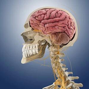
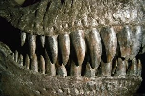
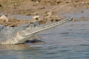
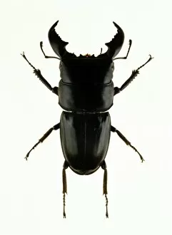
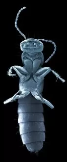
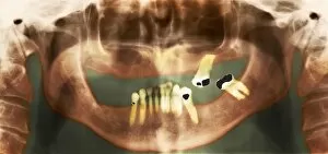
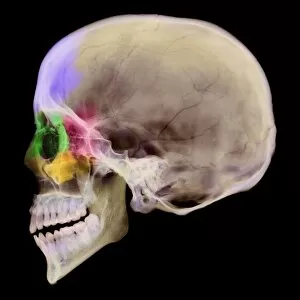
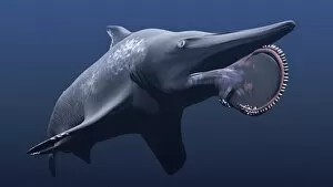
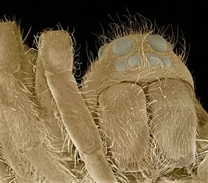
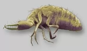
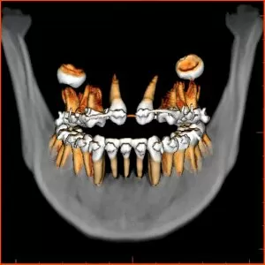
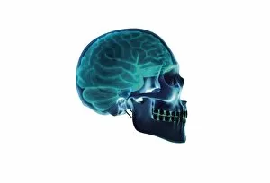
"Jaws: Exploring the Fascinating World of Teeth and Jaws" Discover the intricate world through captivating images that showcase the diversity and power of these remarkable structures. From Leonardo da Vinci's detailed sketches of skull anatomy to breathtaking underwater shots, this collection takes you on a journey into the realm of teeth. Witness the awe-inspiring sight of a Whale Shark with its mouth wide open, gracefully feeding in Australian waters. This gentle giant reminds us that even massive creatures rely on their impressive jaws for sustenance. Delve into the depths alongside a Great White Shark as it glides effortlessly through South Australia's crystal-clear waters. A close-up view reveals its menacing head and formidable open mouth, reminding us why Carcharodon carcharias is known as one of nature's most fearsome predators. Contrasting with these powerful marine creatures, we explore other fascinating jaw structures. The delicate yet sturdy horse skull serves as a reminder that different species have evolved unique adaptations for survival. Take a peek inside our own mouths with panoramic dental X-rays, showcasing how our teeth fit together like puzzle pieces to aid in chewing and speaking. These intricate arrangements are mirrored by another close-up shot capturing every detail of a Great White Shark's toothy grin - an intimidating display perfected over millions of years. Venturing beyond sharks, witness the incredible regenerative abilities found in Greater Spotted Dogfish teeth and jaws from both North Sea and Mediterranean habitats. Continuously replaced from behind, these sharp dentitions ensure efficient hunting for this lesser-known predator. But it's not just animals who possess impressive jaws; Sumatran Tigers also make an appearance with their mouths agape - displaying their strength while emitting fierce roars across their native lands. Lastly, we peer beneath human skin using X-ray technology to reveal our own skeletal marvels - including intricately structured skulls housing our very own set of pearly whites.