
Jean Baptiste Marc Bourgery Collection
Jean Baptiste Marc Bourgery was a renowned anatomist and artist who made significant contributions to the field of medical illustration in the 19th century


For sale as Licensed Images
Choose your image, Select your licence and Download the media
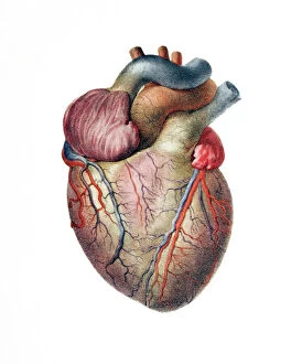
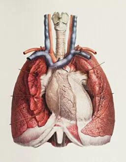
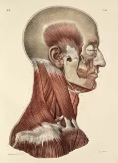
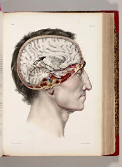
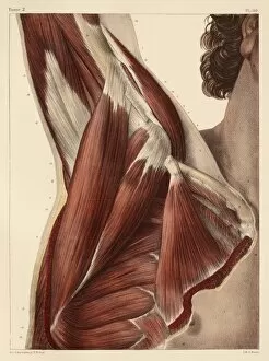
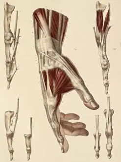
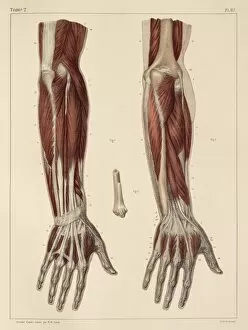
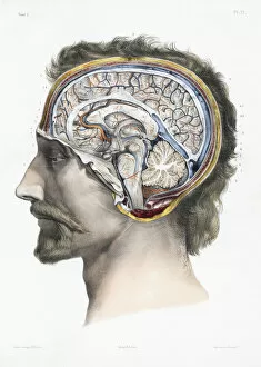
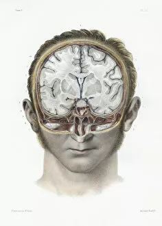
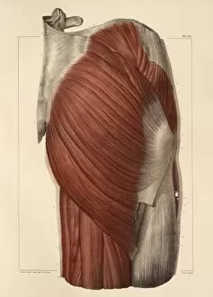
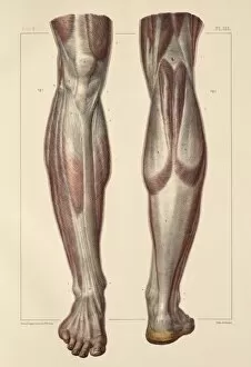
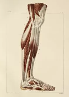
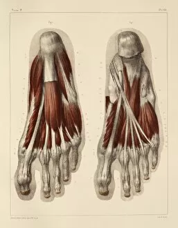
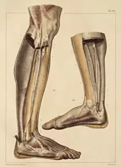
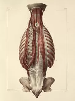
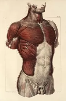
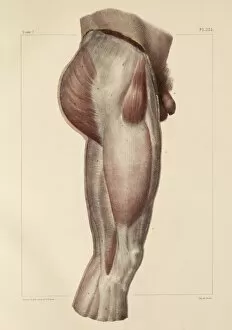
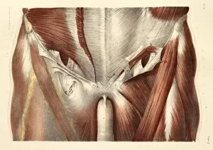
Jean Baptiste Marc Bourgery was a renowned anatomist and artist who made significant contributions to the field of medical illustration in the 19th century. His detailed artwork captured the intricate beauty of human anatomy, showcasing various body parts and systems. One of his notable works is an exquisite depiction of the heart, highlighting its importance as a vital organ that pumps life-giving blood throughout our bodies. In another stunning artwork, Bourgery focused on the complex network of blood vessels within the brain, revealing their crucial role in supplying oxygen and nutrients to this remarkable organ. Bourgery's artistic talent extended beyond internal organs; he also explored the intricacies of head and neck muscles. Through his meticulous brushstrokes, he brought these muscles to life on canvas, showcasing their strength and functionality. In addition to portraying facial muscles with great precision, Bourgery delved into hand muscle anatomy as well. His hand-colored lithograph provided a detailed view of dissected hands, offering valuable insights into how these intricate structures enable us to perform delicate tasks with dexterity. The artist's dedication to capturing every aspect of human anatomy is evident in his depictions of forearm muscles. With careful attention to detail, Bourgery illustrated each muscle group responsible for flexing or extending our forearms—a testament to his commitment towards providing comprehensive anatomical knowledge. Bourgery's work wasn't limited solely to illustrations; he also collaborated with Nicolas-Henri Jacob on volumes dedicated entirely to brain anatomy. These volumes featured sagittal and vertical sections that showcased different perspectives on this enigmatic organ—shedding light on its complexity while captivating viewers with their artistic finesse. Furthermore, Bourgery explored other regions such as pelvic-femoral muscles—an area crucial for movement—and armpit muscles which contribute significantly during upper body activities like lifting or throwing objects.