Joint Collection (#43)

"Exploring the intricate world of joints: from normal knees to X-ray revelations

Background imageJoint Collection: Pelvis bones, artwork

Pelvis bones, artwork

Background imageJoint Collection: Upper body bones, artwork

Upper body bones, artwork

Background imageJoint Collection: Arthrosis of the knee, X-ray

Arthrosis of the knee, X-ray
Arthrosis of the knee, coloured frontal X-ray. The joint is swollen and there are osteophytes, abnormal bony growths (pointed protrusions), seen on the bone at centre

Background imageJoint Collection: Normal lower leg, X-ray

Normal lower leg, X-ray
Normal lower leg, coloured frontal X-ray. The tibia (shinbone, left) and the smaller fibula (centre) articulate with the foot bones (bottom) to form the ankle joint

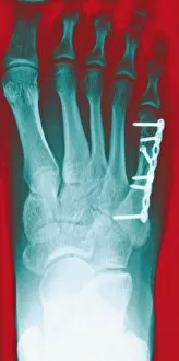
Background imageJoint Collection: Pinned foot bone fracture, X-ray

Pinned foot bone fracture, X-ray
Pinned foot bone fracture. Coloured X-rays of the right foot, showing a metal plate and screws (white) in the foot bone beneath the little toe (upper right)

Background imageJoint Collection: Temporary ankle immobilisation, X-ray

Temporary ankle immobilisation, X-ray
Temporary ankle immobilisation, coloured profile X-ray. These long pins (white rods) are holding the bone fragments in position until they can be operated on and reset properly

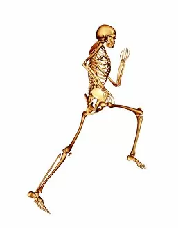
Background imageJoint Collection: Skeleton running

Skeleton running, computer artwork

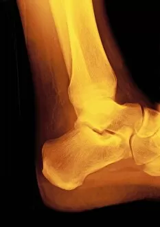
Background imageJoint Collection: Normal ankle joint, X-ray

Normal ankle joint, X-ray
Normal ankle (talocrural) joint, coloured profile X-ray. The front of the foot is at right, with the heel at lower centre

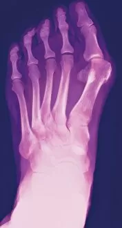
Background imageJoint Collection: Bunion, X-ray

Bunion, X-ray
Bunion. Coloured X-ray of a bunion (hallux valgus, upper right), the swelling of the joint between the big toe and the first metatarsal bone in the foot

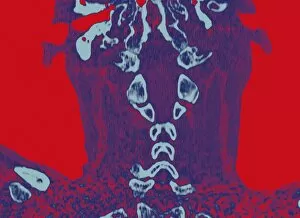
Background imageJoint Collection: Normal neck, CT scan

Normal neck, CT scan
Normal neck, coloured coronal CT (computed tomography) scan. Spinal bones (cervical vertebrae) are light blue, and soft tissue is dark blue. These bones make up the top part of the spine

Background imageJoint Collection: Anterior cruciate ligament repair, X-ray

Anterior cruciate ligament repair, X-ray
Anterior cruciate ligament repair. Coloured profile X-ray of fixation devices (white) in the bones of the knee, used to hold the anterior cruciate ligament (not seen) in place

Background imageJoint Collection: Rheumatoid arthritis

Rheumatoid arthritis
MODEL RELEASED. Rheumatoid arthritis. General practitioner examining a patients hand for signs of rheumatoid arthritis. This condition is caused by the immune system attacking the bodys own tissues

Background imageJoint Collection: Prosthetic ball joint

Prosthetic ball joint

Background imageJoint Collection: Syndecan-4 molecule

Syndecan-4 molecule
Molecular stick reprensation of the syndecan-4 molecule. It plays an important role in the cartilage breakdown in osteoarthritis

Background imageJoint Collection: Ribcage, computer artwork

Ribcage, computer artwork
Ribcage. Computer artwork of the pectoral girdle and ribcage as seen from the front

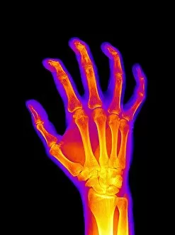
Background imageJoint Collection: Arthritic hand, X-ray

Arthritic hand, X-ray
Arthritic hand, coloured X-ray

Background imageJoint Collection: Topex-Poseidon satellite, artwork

Topex-Poseidon satellite, artwork

Background imageJoint Collection: Arthritic hand, X-ray artwork

Arthritic hand, X-ray artwork
Arthritic hand. Computer artwork of an X-ray of a hand affected by rheumatoid arthritis

Background imageJoint Collection: Jason-2 satellite, artwork

Jason-2 satellite, artwork

Background imageJoint Collection: Jason-1 satellite, artwork

Jason-1 satellite, artwork

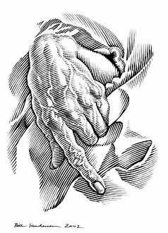
Background imageJoint Collection: Aged hand, artwork

Aged hand, artwork

Background imageJoint Collection: Bluebottle fly foot, SEM

Bluebottle fly foot, SEM
Bluebottle fly foot, coloured scanning electron micrograph (SEM). Magnification: x250 when printed at 10 centimetres wide

Background imageJoint Collection: Oceanography satellites, artwork

Oceanography satellites, artwork
Oceanography satellites orbiting the Earth, artwork. They are Topex-Poseidon (left) and Jason-1 (right). Topex-Poseidon, launched in 1992

Background imageJoint Collection: Osteoarthritic knee, artwork

Osteoarthritic knee, artwork
Artwork of bones of the knee joint: femur (thigh bone, at top) and tibia (shin bone, at bottom) showing osteoarthritis stage 4

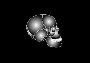
Background imageJoint Collection: Human skull anatomy, artwork

Human skull anatomy, artwork
Human skull anatomy. Artwork showing the structure and anatomy of a human skull, including the sutures (jagged lines), the joints between the fused skull bones

Background imageJoint Collection: Arthritic hands

Arthritic hands
MODEL RELEASED. Arthritic hands. Hands of a person that have become deformed due to arthritis. Arthritis is a degenerative condition that affects the joints causing inflammation and pain

Background imageJoint Collection: Replacement ankle joint, artwork

Replacement ankle joint, artwork
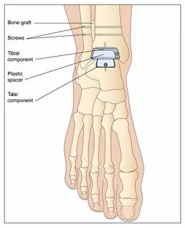
Replacement ankle joint. Artwork showing the location of a prosthetic ankle joint. This device is used to replace a worn or damaged joint

Background imageJoint Collection: Shoulder bones, artwork

Shoulder bones, artwork
Shoulder bones. Artwork showing the bones of the shoulder in profile. The large triangular bone at left is the shoulder blade (scapular)

Background imageJoint Collection: Hip joint, artwork

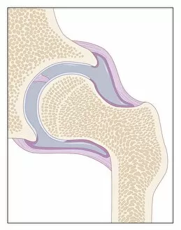
Hip joint, artwork. This is a ball and socket joint. The socket is a cup-shaped cavity in the pelvic bone (upper left), which the ball (centre left), at the head of the femur (thigh bone)

Background imageJoint Collection: Elbow replacement, artwork

Elbow replacement, artwork
Elbow replacement. Artwork showing a prosthetic elbow joint in place. The elbow joint is formed by the articulation of the humerus (upper arm bone) and the ulna and radius (lower arm bones)

Background imageJoint Collection: Female pelvis, artwork

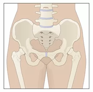
Female pelvis, artwork. The lowest lumbar vertebra of the spinal column (top centre) forms a joint with the sacrum (triangular)

Background imageJoint Collection: Anterior cruciate ligament tear, CT scan

Anterior cruciate ligament tear, CT scan
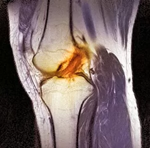
Anterior cruciate ligament tear. Coloured computer tomography (CT) scan of the knee joint of a patient with a tear of the anterior cruciate ligament (ACL)

Background imageJoint Collection: Knee replacement alignment, artwork

Knee replacement alignment, artwork
Knee replacement alignment. Artwork showing an instrument being used to determine the correct positioning of a partial knee replacement

Background imageJoint Collection: Fuel poverty

Fuel poverty
MODEL RELEASED. Fuel poverty. Elderly womans hands holding a fuel bill, representing fuel poverty. She is 90 years old. The elderly often live on reduced incomes

Background imageJoint Collection: Hand anatomy, artwork

Hand anatomy, artwork
hand, bone, skeleton

Background imageJoint Collection: Partial knee replacement, artwork

Partial knee replacement, artwork
Partial knee replacement. Artwork showing a partial knee replacement being fitted. A partial knee replacement is carried out when only one side of the knee is damaged or diseased

Background imageJoint Collection: Bone death, artwork

Bone death, artwork
Bone death. Computer artwork of a human hip and knee joint affected by osteonecrosis - death of the bone tissue. This condition is caused by an impaired blood supply to the bone cells

Background imageJoint Collection: Broken hip, X-ray

Broken hip, X-ray

Background imageJoint Collection: Arteritis of the knee, X-ray

Arteritis of the knee, X-ray
Arteritis of the knee. Coloured X-ray image of the knee of an 85-year-old patient with arteritis. Arteritis is an inflammation of the walls of arteries

Background imageJoint Collection: Fractured elbow, CT scan

Fractured elbow, CT scan
Fractured elbow. Coloured 3D computed tomography (CT) scan of the elbow of an 84-year-old patient that was fractured after a fall

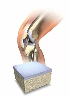
Background imageJoint Collection: Normal human knee, artwork

Normal human knee, artwork
Normal human knee. Artwork showing the internal anatomy of a healthy human knee, with an inset (bottom) of the cartilage (grey) that covers the ends of the bones forming this joint

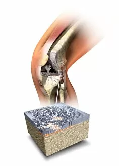
Background imageJoint Collection: Damaged knee, artwork

Damaged knee, artwork
Damaged knee. Artwork showing the internal anatomy of a damaged human knee, with an inset (bottom) of the cartilage (grey) that covers the ends of the bones forming this joint

Background imageJoint Collection: Osteoporosis, artwork

Osteoporosis, artwork
Osteoporosis. Artwork of vertical sections comparing the upper parts of a healthy femur (left) and one with reduced bone mass (right)

Background imageJoint Collection: Shrapnel injury, X-ray

Shrapnel injury, X-ray

Background imageJoint Collection: Arteritis, 3d CT scan

Arteritis, 3d CT scan
Arteritis. Coloured 3d computed tomography (CT) angiogram of the abdominal aorta in a 60-year-old patient after treatment for arteritis

Background imageJoint Collection: Charcot arthropathy, MRI scans

Charcot arthropathy, MRI scans
Charcot arthropathy. Coloured magnetic resonance imaging (MRI) scans of the foot of a 33-year-old patient with Charcot arthropathy

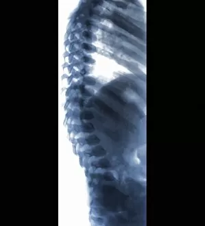
Background imageJoint Collection: Spine in Hurler Syndrome, X-ray

Spine in Hurler Syndrome, X-ray
Spine in Hurler Syndrome. Coloured X-ray of the spine of a 26-month-old child with Hurler syndrome (also called mucopolysaccharidosis type I, MPS I)

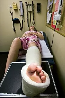
Background imageJoint Collection: Woman with a broken ankle

Woman with a broken ankle
MODEL RELEASED. Woman with a broken ankle waiting in a doctors office. A cast around her lower leg, ankle and foot has immobilised the joint, allowing it to heal


For sale as Licensed Images

Choose your image, Select your licence and Download the media

"Exploring the intricate world of joints: from normal knees to X-ray revelations. " Witness the marvels of our body's architecture as we delve into the anatomy of a human knee joint, where strength and flexibility meet. Step into Shoreham-by-Sea's bustling High Street and discover a hidden gem – the Butchers Shop, where skilled hands expertly carve beef joints for your culinary delight. Unveiling Marie Curie's groundbreaking discoveries in science, she proved that knowledge is indeed a powerful joint between passion and dedication. The Vegetabull proudly presents "The Joint Poster, " showcasing an array of vibrant vegetables coming together harmoniously to create delicious meals bursting with flavor. Market Reports reveal how English Country Squires skillfully carve beef joints, ensuring every slice is perfection on a plate fit for royalty. Hip replacement becomes art as talented artists capture its essence through stunning artwork, reminding us that even in adversity, beauty can emerge. Peering through an X-ray lens reveals the wonders of modern medicine - witness the transformation from pain to mobility with a hip joint replacement. Delve into nature's microcosm as we explore an ant under scanning electron microscopy (SEM), highlighting their remarkable ability to navigate their tiny yet complex joints effortlessly. A closer look at bunions through X-ray imagery reminds us that even small imperfections can impact our daily lives; seek proper care and relief when needed. Shoulder tendinitis captured by X-ray serves as a reminder to nurture our bodies while pushing boundaries – listen closely when they whisper about overuse or strain.