
Joint Collection (#7)

"Exploring the intricate world of joints: from normal knees to X-ray revelations

Background imageJoint Collection: Intercostal nerves anatomy engraving 1866

Intercostal nerves anatomy engraving 1866
Atlas d anatomie descriptive du corps humain C. Bonamy - Paul Broca Victor Masson et Fils Paris 1866

Background imageJoint Collection: Illustration, ellipsoidal joint, arrows indicating possible directions of movement

Illustration, ellipsoidal joint, arrows indicating possible directions of movement

Background imageJoint Collection: Illustration, pivot joint, arrows indicating possible directions of movement

Illustration, pivot joint, arrows indicating possible directions of movement

Background imageJoint Collection: Anatomy of hip fracture

Anatomy of hip fracture

Background imageJoint Collection: Human knee cutaway illustration

Human knee cutaway illustration
Cutaway illustration of human knee showing detailed side and front views

Background imageJoint Collection: Temporomandibular joint, normal and dislocated

Temporomandibular joint, normal and dislocated

Background imageJoint Collection: Anatomy of a swollen foot

Anatomy of a swollen foot

Background imageJoint Collection: Dandelion -Taraxacum- growing out of the joint of a concrete staircase

Dandelion -Taraxacum- growing out of the joint of a concrete staircase

Background imageJoint Collection: MITERING TOOL, 1867. Diagram of a mitering tool. Engraving, American, 1867

MITERING TOOL, 1867. Diagram of a mitering tool. Engraving, American, 1867

Background imageJoint Collection: Photo of Generic 1930s Juke Joint

Photo of Generic 1930s Juke Joint
UNSPECIFIED - CIRCA 1930: Photo of Generic 1930s Juke Joint (Photo by Michael Ochs Archives/Getty Images)

Background imageJoint Collection: Franco-British Exhibition - Marianne, England and Britannia

Franco-British Exhibition - Marianne, England and Britannia
Franco-British Exhibition, London, 1908 - Marianne, A personification of England (a Medieval Queen) and Britannia meet below their respective flags

Background imageJoint Collection: Human hand musculature, artwork F007 / 3436

Human hand musculature, artwork F007 / 3436
Human hand musculature, computer artwork

Background imageJoint Collection: Arthrosis of the hand, X-ray F006 / 4616

Arthrosis of the hand, X-ray F006 / 4616
Coloured X-ray of the hand of a patient with arthrosis, particularly the saddle joint. Arthrosis is a degenerative joint disease that damages cartilage and bone

Background imageJoint Collection: Human arm musculature, artwork F007 / 5764

Human arm musculature, artwork F007 / 5764
Human arm musculature, computer artwork

Background imageJoint Collection: X-ray machine in foot surgery

X-ray machine in foot surgery. Surgeons using a portable operating theatre X-ray machine to check the position of pins that have been inserted into a patients foot bones during surgery

Background imageJoint Collection: Human leg bones, artwork F007 / 2546

Human leg bones, artwork F007 / 2546
Human leg bones, computer artwork

Background imageJoint Collection: Human foot bones, artwork F007 / 1264

Human foot bones, artwork F007 / 1264
Human foot bones, computer artwork

Background imageJoint Collection: Human leg bones, artwork F007 / 1256

Human leg bones, artwork F007 / 1256
Human leg bones, computer artwork

Background imageJoint Collection: Human arm bones, artwork F007 / 1064

Human arm bones, artwork F007 / 1064
Human arm bones, computer artwork

Background imageJoint Collection: Hip joint bones and anatomy, artwork C014 / 2032

Hip joint bones and anatomy, artwork C014 / 2032
Hip joint bones and anatomy. Artwork of the bones that form the two hip joints (right and left). The hip is a ball-and-socket joint that provides the mobility needed for walking and running

Background imageJoint Collection: Radiologist examining knee MRI scans C014 / 1300

Radiologist examining knee MRI scans C014 / 1300
MODEL RELEASED. Radiologist examining knee MRI (magnetic resonance imaging) scans

Background imageJoint Collection: Dislocated wrist, X-ray C017 / 7565

Dislocated wrist, X-ray C017 / 7565
Dislocated wrist. X-ray of the wrist of a 16 year old male that has been dislocated

Background imageJoint Collection: Fist clenching movement, artwork C016 / 6795

Fist clenching movement, artwork C016 / 6795
Fist clenching movement. Artwork of the muscles of the forearm and hand from the front, with red arrows showing the direction of movement of the fingers during flexion to form a fist

Background imageJoint Collection: Right shoulder and nerve plexus, artwork C016 / 6811

Right shoulder and nerve plexus, artwork C016 / 6811
Right shoulder and nerve plexus. Artwork of the nerves (yellow), arteries (red), and bones of the right shoulder, seen from the front

Background imageJoint Collection: Little and ring finger flexion, artwork C016 / 6793

Little and ring finger flexion, artwork C016 / 6793
Little and ring finger flexion. Artwork of the muscles of the forearm and hand from the front, with red arrows showing the direction of movement of the little and ring (fourth and third)

Background imageJoint Collection: Healthy knee, CT scan C018 / 0413

Healthy knee, CT scan C018 / 0413
Healthy knee. Coloured frontal computed tomography (CT) scan projected on to a magnetic resonance imaging (MRI) scan of the knee of a 30 year old

Background imageJoint Collection: Paprosky femur defect classification C016 / 6621

Paprosky femur defect classification C016 / 6621
Paprosky femur defect classification. Diagram showing the classification system for femur cortex defects used to assess revision (replacement or repair) of a hip implant

Background imageJoint Collection: Prosthetic knees and obesity, X-ray C016 / 6598

Prosthetic knees and obesity, X-ray C016 / 6598
Prosthetic knees and obesity. Coloured X-ray of prosthetic knee joints (white) in an obese patient. This patient weighs 102 kilograms

Background imageJoint Collection: Prosthetic knees and obesity, X-ray C016 / 6596

Prosthetic knees and obesity, X-ray C016 / 6596
Prosthetic knees and obesity. X-ray of prosthetic knee joints (white) in an obese patient. This patient weighs 102 kilograms

Background imageJoint Collection: Arm anatomy, artwork C013 / 4583

Arm anatomy, artwork C013 / 4583
Arm anatomy. Computer artwork of an oblique anterior view looking up a human arm, showing muscles and tendons (brown and white), bones and ligaments (white), arteries and veins (red and blue)

Background imageJoint Collection: Buttock muscles, artwork C013 / 4414

Buttock muscles, artwork C013 / 4414
Buttock muscles. Computer artwork of a posterior view of the hips showing some of the muscles (red) of the buttocks. These are shown attached to the femurs (thigh bones) and the pelvis bones

Background imageJoint Collection: Lorius garrulus, chattering lory

Lorius garrulus, chattering lory
Plate 44, a watercolour from the John Reeves Collection of Zoological Drawings from Canton, China

Background imageJoint Collection: Fractured foot, coloured X-ray

Fractured foot, coloured X-ray
Fractured foot. Coloured X-rays of a fractured fifth metatarsal (to the right of each image). The fracture is seen as a small black crack

Background imageJoint Collection: Ankle joint, X-ray

Ankle joint, X-ray
Ankle joint. X-ray of the lateral view of a normal ankle joint, or talocrural joint, on a twenty-four year old male

Background imageJoint Collection: Knee bones

Knee bones
Knee joint. Computer artwork of a healthy knee joint, seen from the side. The knee is the largest joint in the body. It is formed by the articulation of the femur (thigh bone)

Background imageJoint Collection: Broken shoulder, X-ray

Broken shoulder, X-ray
Broken shoulder. X-ray of the shoulder of a patient with a fracture in the neck of the humerus (upper arm bone)

Background imageJoint Collection: Human skull anatomy, diagram

Human skull anatomy, diagram
Human skull anatomy. Diagram showing the structure and anatomy of a human skull, including the sutures (jagged lines), the joints between the fused skull bones

Background imageJoint Collection: Elbow anatomy, artwork

Elbow anatomy, artwork. The elbow joint is formed by the articulation of the humerus (upper arm bone) and the ulna and radius (lower arm bones)

Background imageJoint Collection: Torn ankle ligaments, artwork

Torn ankle ligaments, artwork. Ligaments are bands of fibrous connective tissue that connect bones to other bones. This type of injury, commonly known as a sprained ankle

Background imageJoint Collection: Ant, SEM

Ant, SEM
Ant (family Formicidae), coloured scanning electron micrograph (SEM). One of its compound eyes (black) is seen, as well as two long antennae that are mounted on the head between the eyes

Background imageJoint Collection: Duchess of Albany with Charles Edward

Duchess of Albany with Charles Edward
Princess Helena of Waldeck-Pyrmont, Duchess of Albany with her son, Prince Charles Edward of Albany (1884-1954). Charlie was born posthumously

Background imageJoint Collection: Human arm bones, shoulder blade, humrus leading to elbow joint, ulna and radius extending

Human arm bones, shoulder blade, humrus leading to elbow joint, ulna and radius extending to wrist bones, hand and fingers, side view

Background imageJoint Collection: Studies of the Head and Shoulders of a Man, c1480 (1945). Artist: Leonardo da Vinci

Studies of the Head and Shoulders of a Man, c1480 (1945). Artist: Leonardo da Vinci
Studies of the Head and Shoulders of a Man, c1480 (1945). From The Drawings of Leonardo da Vinci. [Reynal & Hitchcock, New York, 1945]

Background imageJoint Collection: Dissection of a Bears Foot to the Right, c1480 (1945). Artist: Leonardo da Vinci

Dissection of a Bears Foot to the Right, c1480 (1945). Artist: Leonardo da Vinci
Dissection of a Bears Foot to the Right, c1480 (1945). From The Drawings of Leonardo da Vinci. [Reynal & Hitchcock, New York, 1945]

Background imageJoint Collection: Measured Drawing of the Near Fore-Leg of a Horse, c1480 (1945). Artist: Leonardo da Vinci

Measured Drawing of the Near Fore-Leg of a Horse, c1480 (1945). Artist: Leonardo da Vinci
Measured Drawing of the Near Fore-Leg of a Horse, c1480 (1945). From The Drawings of Leonardo da Vinci. [Reynal & Hitchcock, New York, 1945]

Background imageJoint Collection: Studies of Horses Fore-Legs and Measured Drawings of Horses Heads, c1480 (1945)

Studies of Horses Fore-Legs and Measured Drawings of Horses Heads, c1480 (1945). Artist: Leonardo da Vinci
Studies of Horses Fore-Legs and Measured Drawings of Horses Heads, c1480 (1945). From The Drawings of Leonardo da Vinci. [Reynal & Hitchcock, New York, 1945]

Background imageJoint Collection: Four Studies of Horses Legs, c1480 (1945). Artist: Leonardo da Vinci

Four Studies of Horses Legs, c1480 (1945). Artist: Leonardo da Vinci
Four Studies of Horses Legs, c1480 (1945). From The Drawings of Leonardo da Vinci. [Reynal & Hitchcock, New York, 1945]

Background imageJoint Collection: Sir Benjamin Brodie, English doctor, c1860 (1878)

Sir Benjamin Brodie, English doctor, c1860 (1878). Benjamin Collins Brodie (1783-1862) was a physiologist and surgeon who pioneered research into bone and joint disease


For sale as Licensed Images

Choose your image, Select your licence and Download the media

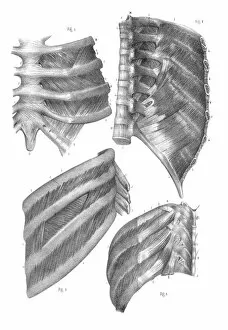
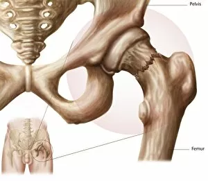
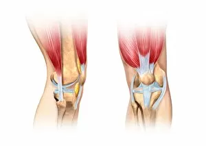
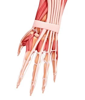
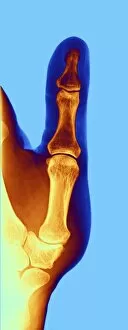
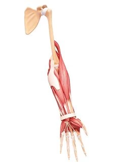
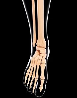
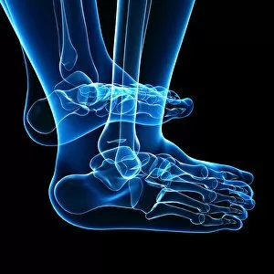
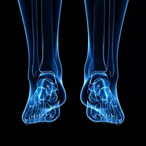
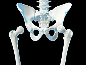
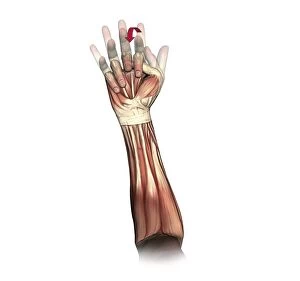
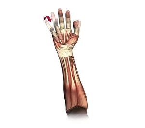
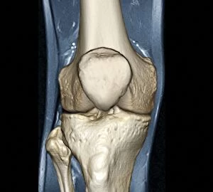
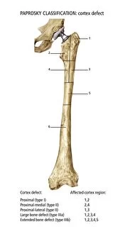
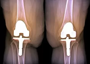
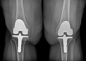
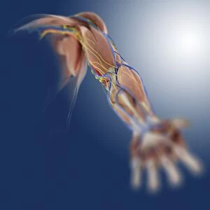
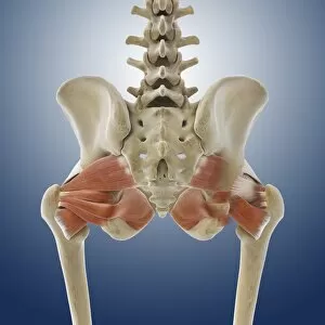
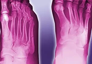
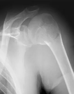
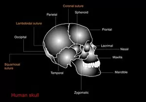
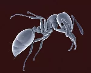
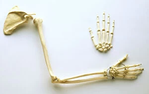
"Exploring the intricate world of joints: from normal knees to X-ray revelations. " Witness the marvels of our body's architecture as we delve into the anatomy of a human knee joint, where strength and flexibility meet. Step into Shoreham-by-Sea's bustling High Street and discover a hidden gem – the Butchers Shop, where skilled hands expertly carve beef joints for your culinary delight. Unveiling Marie Curie's groundbreaking discoveries in science, she proved that knowledge is indeed a powerful joint between passion and dedication. The Vegetabull proudly presents "The Joint Poster, " showcasing an array of vibrant vegetables coming together harmoniously to create delicious meals bursting with flavor. Market Reports reveal how English Country Squires skillfully carve beef joints, ensuring every slice is perfection on a plate fit for royalty. Hip replacement becomes art as talented artists capture its essence through stunning artwork, reminding us that even in adversity, beauty can emerge. Peering through an X-ray lens reveals the wonders of modern medicine - witness the transformation from pain to mobility with a hip joint replacement. Delve into nature's microcosm as we explore an ant under scanning electron microscopy (SEM), highlighting their remarkable ability to navigate their tiny yet complex joints effortlessly. A closer look at bunions through X-ray imagery reminds us that even small imperfections can impact our daily lives; seek proper care and relief when needed. Shoulder tendinitis captured by X-ray serves as a reminder to nurture our bodies while pushing boundaries – listen closely when they whisper about overuse or strain.