
Kidneys Collection (#3)
"Exploring the intricate world of kidneys: a journey through art and science" Embarking on a full body scan


For sale as Licensed Images
Choose your image, Select your licence and Download the media
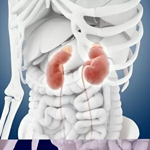
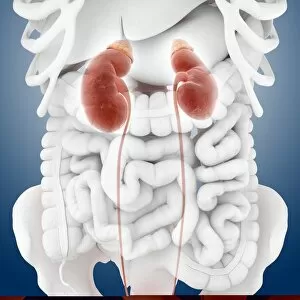
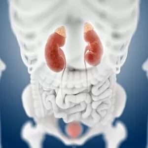
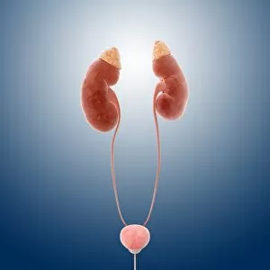
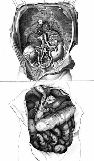
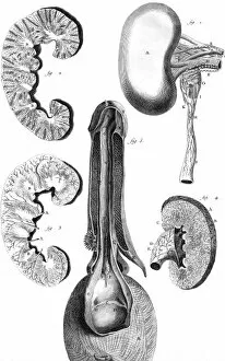
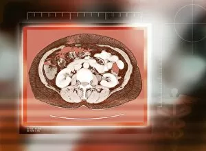
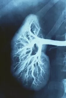
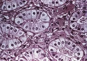
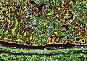
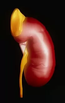
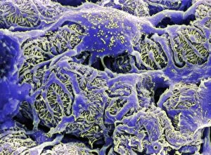
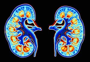
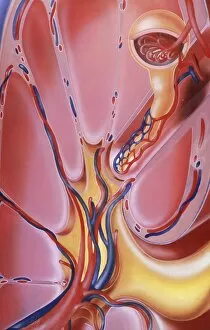
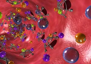
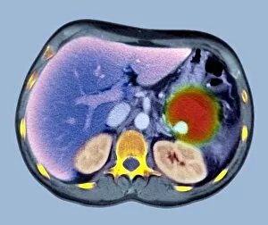
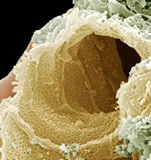
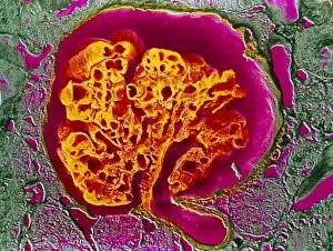
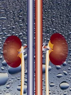
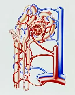
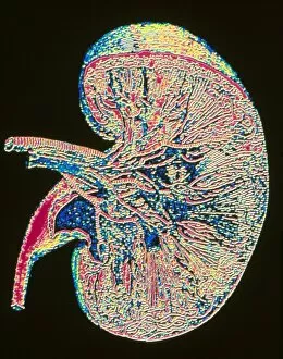
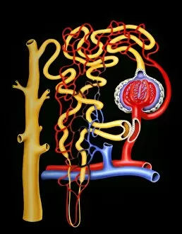
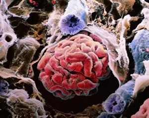
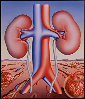
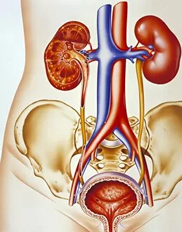
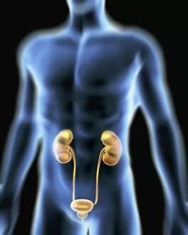
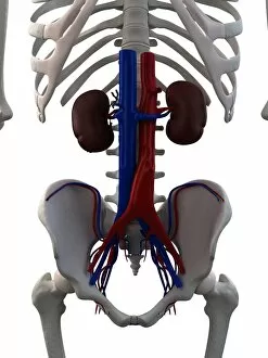
"Exploring the intricate world of kidneys: a journey through art and science" Embarking on a full body scan, we delve into the depths of our anatomy to uncover the secrets held within. The MRI scan reveals the complex network of our cardiovascular system, intertwining with historical artwork that depicts its significance throughout time. Zooming in further, we encounter kidney tubules in section - tiny structures responsible for filtering waste from our blood. A vivid colour MRI scan showcases the exquisite placement of these vital organs alongside the liver in our abdomen. Did you know? We actually possess spare kidneys. Just like pig anatomy depicted in ancient artwork, humans have two kidneys but can function with just one if needed. Our bodies are truly remarkable. Taking an even closer look, we witness the mesmerizing beauty of kidney glomeruli under scanning electron microscopy (SEM). These intricate structures play a crucial role in filtration and maintaining fluid balance within our bodies. As we explore deeper into our venous system, we marvel at its complexity and interconnectedness. X-ray images reveal abdominal arteries pulsating with life - nourishing every corner of our being. Returning to SEM imagery once again, we observe the delicate web of blood vessels surrounding each kidney glomerulus, and is here that filtration occurs, ensuring waste products are efficiently removed from our bloodstream. In this captivating journey through art and science, we gain a newfound appreciation for these incredible organs known as kidneys. They not only filter waste but also regulate electrolyte levels and maintain overall homeostasis within us – silently working day and night to keep us healthy.