
Lateral Collection (#8)
"Lateral: Exploring the Intricacies of Nature's Design" Delving into the intricate network of brain blood vessels, as captured in a mesmerizing 3D angiogram C007 / 1981


For sale as Licensed Images
Choose your image, Select your licence and Download the media
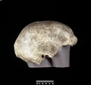
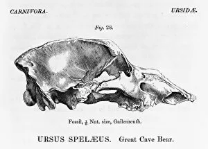
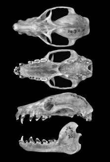
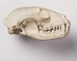
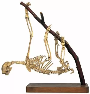
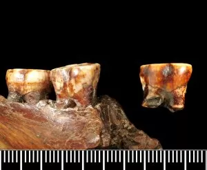
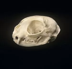
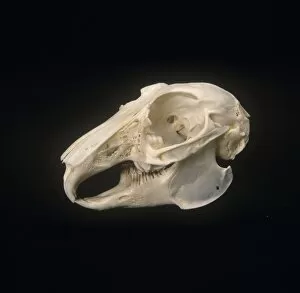
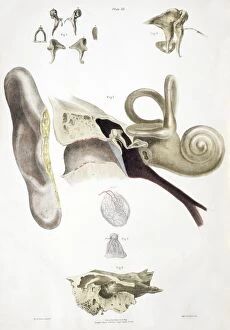
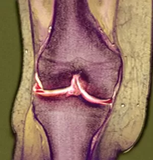
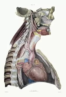
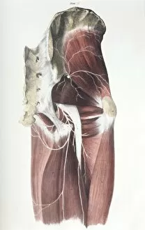
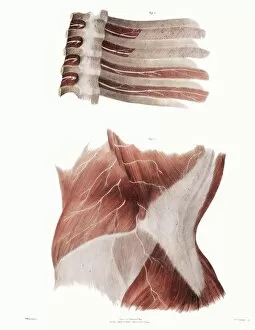
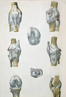
"Lateral: Exploring the Intricacies of Nature's Design" Delving into the intricate network of brain blood vessels, as captured in a mesmerizing 3D angiogram C007 / 1981. Unveiling the hidden wonders of the human knee joint through an anatomy study that reveals its lateral complexities. Witnessing the majestic Eurasian lynx (Lynx lynx) in all its glory, with its captivating lateral movements and stealthy demeanor. Marveling at the delicate intricacy of brain blood vessels, showcasing nature's remarkable lateral pathways within our most vital organ. Transporting ourselves back to 1956 French Grand Prix, where mechanics examine Trintignants Bugatti Type 251 chassis and its straight 8 Bugatti 251 engine from a lateral perspective. Glimpsing at the striking Red-billed Chough (Pyrrhocorax pyrrhocorax) at Alpine Zoo Innsbruck, Tyrol, Austria, Europe - a testament to nature's lateral adaptations for survival in high altitudes. Admiring an exquisite artwork from 1825 depicting male groin arteries - a fascinating exploration into our body's intricate vascular system from a unique lateral viewpoint. Discovering head and neck muscles through an extraordinary artwork dating back to 1831 - offering insights into their complex interplay and role in our daily movements from various angles including laterally. Examining a tonguestone (sharks tooth) adorned with intriguing lateral denticles that enhance these creatures' feeding efficiency and survival strategies over millions of years. Peering into skull anatomy to unravel mysteries hidden beneath our skin; exploring bone structures and cranial features from both frontal and lateral perspectives for comprehensive understanding. Venturing deep within Earth's core as we explore "Earth's Central Fire, " uncovering geological forces that shape our planet's lateral movements and tectonic shifts.