
Longitudinal Collection (#2)

"Exploring the Intricacies of Longitudinal Structures

Background imageLongitudinal Collection: Iroko wood, SEM

Iroko wood, SEM
Iroko wood. Coloured scanning electron micrograph (SEM) of a longitudinal section through Iroko (Chlorophora excelsa) wood

Background imageLongitudinal Collection: Mitosis in root tip of Broad Bean, LM

Mitosis in root tip of Broad Bean, LM
Light micrograph (longitudinal section) of the cells in the root tip of a broad bean plant Vicia faba. The dark spot in the centre of each cell is the nucleus

Background imageLongitudinal Collection: Earthworm, longitudinal section

Earthworm, longitudinal section
Earthworm. Light micrograph of a longitudinal section through the body of a round segmented earthworm (Lumbricus terrestris), showing the first 14 anterior segments

Background imageLongitudinal Collection: Crayfish compound eye, light micrograph

Crayfish compound eye, light micrograph
Crayfish compound eye. Light micrograph of a longitudinal section through the compound eye (top) of a freshwater crayfish (Astacus astacus)

Background imageLongitudinal Collection: Oesophagus wall, light micrograph

Oesophagus wall, light micrograph
Oesophagus wall. Coloured light micrograph of a section through the human oesophagus, which passes food from the mouth to the stomach

Background imageLongitudinal Collection: Trachea epithelium, light micrograph

Trachea epithelium, light micrograph
Trachea epithelium. Light micrograph of a vertical section through the pseudostratified columnar epithelium from the trachea

Background imageLongitudinal Collection: Longitudinal sections, artwork, 1891

Longitudinal sections, artwork, 1891
Longitudinal sections. Historical artwork of a human being (bottom) and other organisms. Illustration by Ernst Haeckel, published in the 1891 edition of his Anthropogenie

Background imageLongitudinal Collection: Blood vessel types, artwork

Blood vessel types, artwork
Blood vessel types. Artworks of the three types of blood vessels, from left: arteries, veins, and capillaries. Arteries are thick blood vessels carrying oxygenated blood around the body away from

Background imageLongitudinal Collection: Ovarian blood vessel, SEM

Ovarian blood vessel, SEM
Ovarian blood vessel. Coloured scanning electron micrograph (SEM) of a section through a blood vessel in an ovary. Red blood cells are seen in the lumen, which is lined with endothelial cells (brown)

Background imageLongitudinal Collection: Micromechanical accelerometer

Micromechanical accelerometer. This tiny acceleration sensor is used in automatic car stability control systems. These systems can sense lateral (sideways)

Background imageLongitudinal Collection: Optic nerve, light micrograph

Optic nerve, light micrograph
Optic nerve. Coloured light micrograph of a section through the optic nerve. The optic nerve (bright pink) collects impulses from the light sensitive cells of the retina and relays them to the brain

Background imageLongitudinal Collection: Eye structure, light micrograph

Eye structure, light micrograph
Eye structure. Coloured light micrograph of a section through the inner surface of a human eye. This slice shows the vitreous humour (transparent liquid in the eye, top), the retina (pink)

Background imageLongitudinal Collection: Brain

Brain, historical artwork. The top drawing shows the brain cut in-between its hemispheres. The front of the brain is on the left

Background imageLongitudinal Collection: Intestine wall, artwork

Intestine wall, artwork
Intestine wall. Computer artwork of a section through the wall of a human intestine, showing its structure. For the labelled diagram see image: C009/1711

Background imageLongitudinal Collection: Small intestine section, light micrograph

Small intestine section, light micrograph
Small intestine section. Light micrograph of a vertical section through the illeum, part of the small intestine. The upper layer (the digestive surface)

Background imageLongitudinal Collection: Kidney, light micrograph

Kidney, light micrograph
Kidney. Light micrograph of a vertical section through a whole kidney. The kidney is surrounded by a capsule (dark purple) under which is the cortex (deep red)

Background imageLongitudinal Collection: Developing frog egg, light micrograph

Developing frog egg, light micrograph
Developing frog egg. Light micrograph of a vertical section through a developing egg laid by a common frog (Rana temporaria temporaria). The egg has reached the blastula stage

Background imageLongitudinal Collection: Stomach section, light micrograph

Stomach section, light micrograph
Stomach section. Light micrograph of a vertical section through a stomach. The upper layer (pink-purple) is the glandular mucosa

Background imageLongitudinal Collection: Skin section, light micrograph

Skin section, light micrograph
Skin section. Semi-polarised light micrograph of a vertical section through human skin. The outer epidermis is the thin layer across top, with the dermis layer beneath it

Background imageLongitudinal Collection: Tongue section, light micrograph

Tongue section, light micrograph
Tongue section. Light micrograph of a vertical section through a mammalian tongue. At top, the surface is covered with circumvallate papillae

Background imageLongitudinal Collection: Kidney cortex, light micrograph

Kidney cortex, light micrograph
Kidney cortex. Light micrograph of a vertical section through the cortex (outer layer) of a kidney. The cortex tissue (across top) is stained a darker red

Background imageLongitudinal Collection: Eukaryotic flagellum structure, artwork

Eukaryotic flagellum structure, artwork
Eukaryotic flagellum structure. Cutaway artwork showing the internal structure of the flagellum in eukaryotic cells. Flagella are tail-like projections used for cell locomotion

Background imageLongitudinal Collection: Intestinal villi anatomy, artwork

Intestinal villi anatomy, artwork
Intestinal villi anatomy. Artwork showing six types of cell found on the surface of villi in the small intestine. Clockwise from upper right they are: enterocytes (green)

Background imageLongitudinal Collection: Slug turning the right way up

Slug turning the right way up. Composite image of a slug righting itself after it was turned on its back (top left). The slugs circular, oblique and longitudinal muscles

Background imageLongitudinal Collection: Clubmoss cone, light micrograph

Clubmoss cone, light micrograph. Longitudinal section through the cone (strobilus) of the clubmoss Lycopodium clavatum. The strobilus consists of a central axis (purple)

Background imageLongitudinal Collection: Fern plant reproduction, light micrograph

Fern plant reproduction, light micrograph. At bottom, the double kidney-shaped gametophyte (prothallus) plant has anchored itself with root-like rhizoids (central black area)

Background imageLongitudinal Collection: Horsetail cone, light micrograph

Horsetail cone, light micrograph. Longitudinal section through a young cone (strobilus) of the rough horsetail (Equisetum hyemale)

Background imageLongitudinal Collection: Liverwort spore capsule, light micrograph

Liverwort spore capsule, light micrograph. Longitudinal section through the thallus and sporangium of a liverwort (Jungammania sp.) The sporangium (spore capsule)

Background imageLongitudinal Collection: Moss reproductive parts, light micrograph

Moss reproductive parts, light micrograph. Longitudinal section through the antheridial cup (male reproductive parts) of a cord moss (Polytrichum commure)

Background imageLongitudinal Collection: Moss spore capsule, light micrograph

Moss spore capsule, light micrograph
Moss spore capsule, polarised light micrograph. Longitudinal section through a spore capsule from a fire moss (Funaria hygrometrica)

Background imageLongitudinal Collection: Pear fruit tissue, light micrograph

Pear fruit tissue, light micrograph
Pear fruit tissue. Light micrograph of a longitudinal section through tissue from the fruit of a pear tree (Pyrus sp.). The section shows stone clusters or sclerieds (red)

Background imageLongitudinal Collection: Shepherds purse seeds, light micrograph

Shepherds purse seeds, light micrograph
Shepherds purse seeds. Light micrograph of a longitudinal section through a fruit of the shepherds purse (Capsell bursa-pastoris), showing eight seeds and embryos (red)

Background imageLongitudinal Collection: Phloem plant cells, light micrograph

Phloem plant cells, light micrograph
Phloem plant cells. Light micrograph of a longitudinal section through phloem tissue in a Cucurbita plant. Most of this tissue is large phloem tubes (stained blue)

Background imageLongitudinal Collection: Leaf abscission layer, light micrograph

Leaf abscission layer, light micrograph
Leaf abscission layer. Light micrograph of a longitudinal section through a dicotyledon stem that has undergone leaf-fall. Part of the abscission layer (orange) is at centre

Background imageLongitudinal Collection: Dandelion flower head, light micrograph

Dandelion flower head, light micrograph
Dandelion flower head. Light micrograph of a longitudinal section through the flower head (capitulum) of a dandelion (Taraxacum officinale)

Background imageLongitudinal Collection: Lilac leaf bud, light micrograph

Lilac leaf bud, light micrograph
Lilac leaf bud. Light micrograph of a cross-section of a bud from a lilac tree (Syringa vulgaris). The bud is comprised of an outer layer of six thick scale leaves (blue and purple)

Background imageLongitudinal Collection: Female yew tree flower, light micrograph

Female yew tree flower, light micrograph
Female yew tree flower. Light micrograph of a longitudinal section through a female flower of the yew tree (Taxus baccata)

Background imageLongitudinal Collection: Huckleberry shoot, light micrograph

Huckleberry shoot, light micrograph. Longitudinal section through the top of a terminal shoot from a huckleberry (Gaylussacia baccata)

Background imageLongitudinal Collection: Abscission layer, light micrograph

Abscission layer, light micrograph
Abscission layer. Light micrograph of a longitudinal section through a stem from the flame nettle (Solenostemon scutellarioides), showing a developing abscission layer (purple, left)

Background imageLongitudinal Collection: Alexander flowers, light micrograph

Alexander flowers, light micrograph
Alexander flowers. Light micrograph of a section through a cluster of alexander flowers (Smyrnium olusatrum). Seen here are cross-sections of the central umbels

Background imageLongitudinal Collection: Shepherds purse seed, light micrograph

Shepherds purse seed, light micrograph
Shepherds purse seed. Light micrograph of a longitudinal section through a seed of the shepherds purse (Capsell bursa-pastoris), showing the embryo in the seed

Background imageLongitudinal Collection: Cucumber flower bud, light micrograph

Cucumber flower bud, light micrograph
Cucumber flower bud. Light micrograph of a longitudinal section through the flower bud of a cucumber (Cucurbita sp.). The female pistil (red)

Background imageLongitudinal Collection: Poppy fruit, light micrograph

Poppy fruit, light micrograph
Poppy fruit. Light micrograph of a longitudinal section through the fruit of an Iceland poppy (Papaver nudicaule), showing the seeds (small dots). The fruit capsule has an outer wall (pericarp)

Background imageLongitudinal Collection: Lung bronchus cross-section, SEM

Lung bronchus cross-section, SEM
Lung bronchus cross-section, coloured scanning electron micrograph (SEM). This longitudinal freeze-fracture has exposed the inner epithelial surface of a bronchus (brown)

Background imageLongitudinal Collection: Liverwort spore case, light micrograph

Liverwort spore case, light micrograph
Liverwort spore case. Light micrograph of a vertical section through a spore case (sporophyte) from a liverwort (Pellia epiphylla)

Background imageLongitudinal Collection: Pine cone scale, light micrograph

Pine cone scale, light micrograph
Pine cone scale. Light micrograph of a vertical section through an ovulate scale from a Scots pine (Pinus sylvestris). This image shows the ovule (oval shape)


For sale as Licensed Images

Choose your image, Select your licence and Download the media

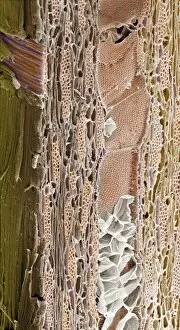
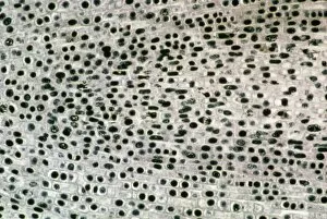
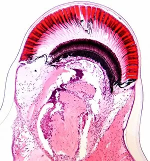
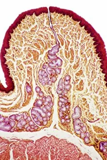
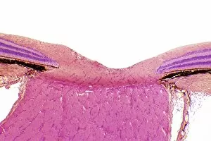
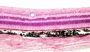
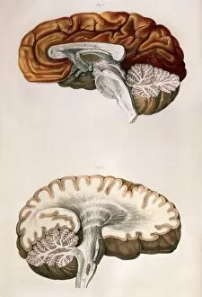
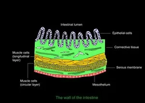
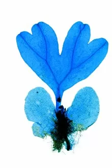
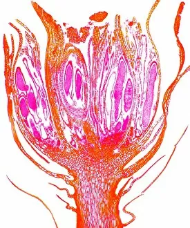
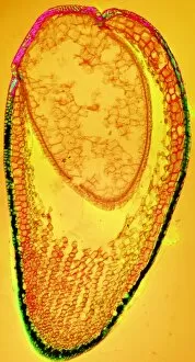
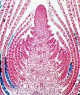
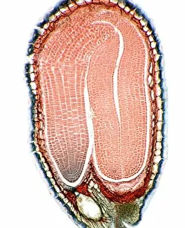
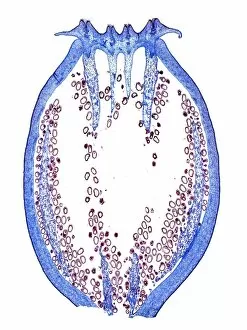
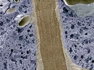
"Exploring the Intricacies of Longitudinal Structures: From Dicotyledon Plant Stems to Jellyfish Artwork" This captivating light micrograph showcases the intricate longitudinal structure of a dicotyledon plant stem, revealing its inner beauty and complexity. Intriguingly artistic, this jellyfish artwork captures the ethereal nature of these creatures with their long tentacles gracefully extending in a longitudinal fashion. A closer look at tendons through SEM reveals their remarkable longitudinal arrangement, highlighting their strength and flexibility in supporting our movements. Delving into the depths of neuroscience, an MRI scan unveils the fascinating longitudinal pathways within a child's brain, offering insights into cognitive development and neural connections. Transporting us back in time, we encounter historical marvels like the Great Eastern steamship. A section view provides a glimpse into its impressive size and design with a prominent longitudinal perspective. The majestic Parthenon of Athens stands as an enduring symbol of ancient Greece's grandeur. Its exquisite columns stretch longitudinally across its magnificent facade, leaving spectators awestruck by its timeless beauty. Across continents to New York City, we find another architectural gem - the Custom House. With its classical design featuring elegant columns arranged longitudinally along its exterior, it exudes both power and grace. Returning to childhood wonderment once more, an MRI scan unravels further mysteries within a child's brain. This mesmerizing image sheds light on how neurological processes unfold longitudinally during crucial developmental stages. Venturing into innovative transportation methods from yesteryears brings us to an atmospheric railway. An elevation view coupled with a detailed longitudinal section illustrates how this ambitious concept aimed to revolutionize travel using air pressure propulsion systems. Unveiling engineering triumphs from history takes us aboard Isambard Kingdom Brunel's visionary creation - The Great Eastern Steam Ship. Despite logistical challenges delaying her launch in 1857, her groundbreaking design showcased through this striking longitudinal section remains a testament to human ingenuity.