Lumbar Collection (#2)

"Lumbar: Exploring the Intricacies of the Human Spine and Abdomen" Delving into the depths of our body's core

Background imageLumbar Collection: Central nervous system with human brain

Central nervous system with human brain

Background imageLumbar Collection: Conceptual image of human backbone

Conceptual image of human backbone

Background imageLumbar Collection: Conceptual image of human brain, skull and spinal cord

Conceptual image of human brain, skull and spinal cord

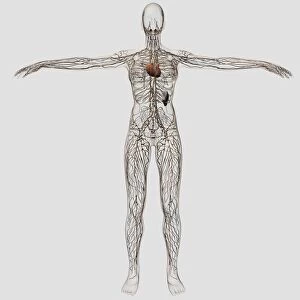
Background imageLumbar Collection: Medical illustration of female circulatory system with heart and veins

Medical illustration of female circulatory system with heart and veins
Medical illustration of full female skeleton standing against white background, three dimensional view

Background imageLumbar Collection: Anatomy of female body with nervous system

Anatomy of female body with nervous system

Background imageLumbar Collection: Illustration of lumbago, lumbar and pelvic regions

Illustration of lumbago, lumbar and pelvic regions
Medicine: lumbago, lumbar and pelvic regions. Drawing

Background imageLumbar Collection: Back pain, artwork F007 / 8390

Back pain, artwork F007 / 8390
Back pain, computer artwork

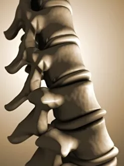
Background imageLumbar Collection: Lumbar spine, artwork F007 / 7357

Lumbar spine, artwork F007 / 7357
Lumbar spine, computer artwork

Background imageLumbar Collection: Lumbar spine, artwork F007 / 7355

Lumbar spine, artwork F007 / 7355
Lumbar spine, computer artwork

Background imageLumbar Collection: Back pain, artwork F007 / 8389

Back pain, artwork F007 / 8389
Back pain, computer artwork

Background imageLumbar Collection: Back pain, artwork F007 / 8387

Back pain, artwork F007 / 8387
Back pain, computer artwork

Background imageLumbar Collection: Back pain, artwork F007 / 8388

Back pain, artwork F007 / 8388
Back pain, computer artwork

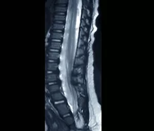
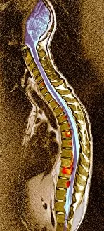
Background imageLumbar Collection: Sciatica, MRI scan

Sciatica, MRI scan
Sciatica. Coloured magnetic resonance imaging (MRI) scan of a sagittal section through the lumbar (lower back) spine of a 35-year-old patient

Background imageLumbar Collection: Spina bifida, MRI scan

Spina bifida, MRI scan
Fibrolipoma of the spine. Magnetic resonance imaging (MRI) scan of a sagittal section through the spine of a 24-month-old patient with spina bifida

Background imageLumbar Collection: Fibrolipoma of the spine, MRI scan

Fibrolipoma of the spine, MRI scan
Fibrolipoma of the spine. Coloured magnetic resonance imaging (MRI) scan of a sagittal section through the thoraco-lumbar spine of a 19-month-old patient

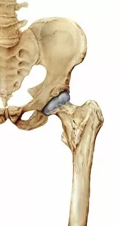
Background imageLumbar Collection: Hip bones, artwork C016 / 7015

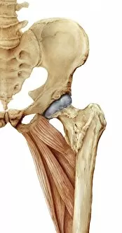
Hip bones, artwork C016 / 7015
Hip bones. Artwork of a frontal (anterior) view of the left hip, showing the bones that articulate to form this joint. At lower right is the femur (thigh bone)

Background imageLumbar Collection: Lumbar spine fusion, artwork C016 / 6993

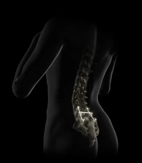
Lumbar spine fusion, artwork C016 / 6993
Lumbar spine fusion. Artwork of pins and screws inserted into a patients lumbar spine in a case of spinal fusion. The pins hold together the patients vertebrae

Background imageLumbar Collection: Hip bones and thigh muscles, artwork C016 / 7016

Hip bones and thigh muscles, artwork C016 / 7016
Hip bones and thigh muscles, artwork

Background imageLumbar Collection: Hip bones, muscles and ligaments, artwork C016 / 7018

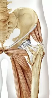
Hip bones, muscles and ligaments, artwork C016 / 7018
Hip bones and thigh muscles. Artwork of a frontal (anterior) view of the left hip, showing bones and some thigh muscles (red). At lower right is the femur (thigh bone)

Background imageLumbar Collection: Scoliosis of the spine, X-ray C016 / 6565

Scoliosis of the spine, X-ray C016 / 6565
Scoliosis of the spine. Coloured X-ray of the chest of a 34 year old female patient with scoliosis (sideways curvature) of the spine

Background imageLumbar Collection: Lower back pain C016 / 9667

Lower back pain C016 / 9667
MODEL RELEASED. Lower back pain

Background imageLumbar Collection: Artificial spinal disc, X-ray C016 / 6561

Artificial spinal disc, X-ray C016 / 6561
Artificial spinal disc. Coloured X-ray of the spine of a 42 year old woman, showing a prosthetic spinal disc (white) in the lumbar (lower) spine

Background imageLumbar Collection: Artificial spinal disc, X-ray C016 / 6562

Artificial spinal disc, X-ray C016 / 6562
Artificial spinal disc. Coloured X-ray of the spine of a 42 year old woman, showing a prosthetic spinal disc (white) in the lumbar (lower) spine

Background imageLumbar Collection: Lower back pain C016 / 9668

Lower back pain C016 / 9668
MODEL RELEASED. Lower back pain

Background imageLumbar Collection: Human torso, coloured mri scan C014 / 2644

Human torso, coloured mri scan C014 / 2644
Normal human torso, coloured frontal magnetic resonance imaging (MRI) scan. The spinal bones (vertebrae) are seen running from the top of the chest to the pelvis

Background imageLumbar Collection: Human spine, anatomical model

Human spine, anatomical model
Human spine. Anatomical model (from 1990) showing the lower (lumbosacral) section of the human spine (backbone)

Background imageLumbar Collection: Pelvis, artwork C014 / 5105

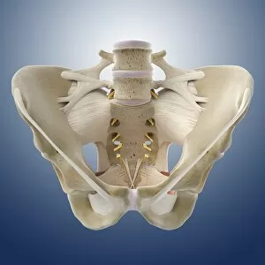
Pelvis, artwork C014 / 5105
Pelvis. Computer artwork of the pelvis showing the ligaments (white) and nerves (yellow). Ligaments are bands of connective tissue that connect bones

Background imageLumbar Collection: Lower back pain, artwork C013 / 5922

Lower back pain, artwork C013 / 5922
Lower back pain, computer artwork

Background imageLumbar Collection: Lower back pain, artwork C013 / 8810

Lower back pain, artwork C013 / 8810
Lower back pain, computer artwork

Background imageLumbar Collection: Coccyx and lower back, X-ray

Coccyx and lower back, X-ray
Coccyx and lower back. Coloured X-ray of a side view of the coccyx and lower back of a healthy adult man. The front of the body is at right

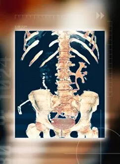
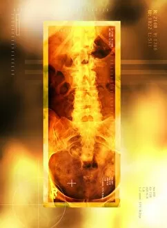
Background imageLumbar Collection: Abdominal bones, 3-D CT scan

Abdominal bones, 3-D CT scan
Abdominal bones, coloured 3-D computer tomography (CT) scan. The lower part of the ribcage is in upper frame, with the pelvis in lower frame. They are connected by the spine, which runs down centre

Background imageLumbar Collection: Spine, computer artwork

Spine, computer artwork
Spine. Computer artwork showing the bones of the spine, shoulder and pelvis on the human body. The spine, or backbone, supports the back and protects the spinal cord

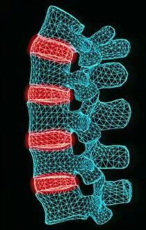
Background imageLumbar Collection: Computer artwork of the five lumbar vertebrae

Computer artwork of the five lumbar vertebrae
Lumbar spine. Computer illustration of the five lumbar vertebrae (blue) of the human spine. The lumbar vertebrae are the largest of the unfused vertebrae in the backbone

Background imageLumbar Collection: Spinal implant

Spinal implant
Spinal column nerve stimulator. Coloured X-ray showing the wires (blue/purple) of a nerve stimulator in the spine of a paraplegic patient

Background imageLumbar Collection: Spondylitis, X-ray

Spondylitis, X-ray
Spondylitis. Coloured X-ray of the lower spine and pelvis of a patient with spondylitis, an inflammation of the synovial joints of the backbone

Background imageLumbar Collection: Potts disease

Potts disease. Coloured X-ray of the spine of a patient with Potts disease, or tuberculosis (TB) of the spine. The vertebrae (brown blocks) have become compacted and inflamed (red, yellow)

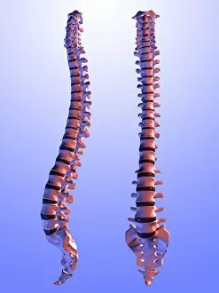
Background imageLumbar Collection: Two views of the human spine

Two views of the human spine. This computer artwork shows a side (lateral) view (left) and an front (anterior) view (right)

Background imageLumbar Collection: Healthy lower back, X-ray

Healthy lower back, X-ray
Healthy lower back, coloured X-ray. The spine runs down centre. It is made up of vertebrae, block- shaped bones that support and protect the spinal cord (not seen)

Background imageLumbar Collection: Coloured X-ray of normal lumbar spine (lower back)

Coloured X-ray of normal lumbar spine (lower back)
Lumbar spine and pelvis. Computer-enhanced X-ray of the healthy lumbar spine (lower back) and pelvis of a human. At top can be seen the lower ribs of the ribcage

Background imageLumbar Collection: Coloured X-ray of normal lower spine and pelvis

Coloured X-ray of normal lower spine and pelvis
Lower spine and pelvis. Coloured X-ray of healthy human lumbar spine (lower back) and pelvic girdle. At top can be seen the 11th and 12th floating ribs on either side of the thoracic vertebrae

Background imageLumbar Collection: Spinal vertebra

Spinal vertebra. Historical artwork of a vertebra (spinal bone) seen from the side. The rear of the spine is at right. This is a lumbar vertebra, one from the lower back

Background imageLumbar Collection: Lumbar spine mobilisation treatment

Lumbar spine mobilisation treatment
MODEL RELEASED. Lumbar spine mobilisation treatment. Physiotherapist manipulating the lumbar (lower) region of a patients spine, during a spinal mobilisation treatment session

Background imageLumbar Collection: Lower back pain, X-ray artwork

Lower back pain, X-ray artwork
Back pain. Computer X-ray artwork of the rear view of the lower back of a human skeleton. The red area represents lower back pain (lumbago)

Background imageLumbar Collection: Back pain, X-ray artwork

Back pain, X-ray artwork
Back pain. Computer X-ray artwork of the rear view of a human skeleton. The red area represents back pain (dorsalgia). Back pain usually originates from the muscles, nerves, bones

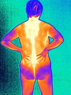
Background imageLumbar Collection: Back pain, thermogram

Back pain, thermogram
Painful back. Thermogram of the back of a naked woman holding her painful back. The colours show variation in temperature

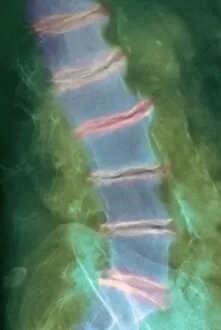
Background imageLumbar Collection: Arthritis of the spine, X-ray

Arthritis of the spine, X-ray

Background imageLumbar Collection: Lower back pain, X-ray

Lower back pain, X-ray
Lower back pain. Conceptual coloured X-ray of the bones of the lower back glowing red in pain. Lower back pain (lumbago) is a very common complaint

Background imageLumbar Collection: Arthritis of the back, X-ray

Arthritis of the back, X-ray
Arthritis of the back, coloured X-ray. Lumbar arthritis caused by chondrocalcinosis, a condition where crystals of calcium pyrophosphate are deposited in soft tissues


For sale as Licensed Images

Choose your image, Select your licence and Download the media

"Lumbar: Exploring the Intricacies of the Human Spine and Abdomen" Delving into the depths of our body's core, a colour MRI scan reveals the intricate beauty of kidneys and liver in vivid detail. Body pain can be debilitating, but through art, we find solace in expressing our struggles and seeking healing for our lumbar region. A coloured X-ray unveils the delicate architecture vertebrae, providing a glimpse into the foundation that supports our spine's strength and flexibility. Behold a normal spine captured by an X-ray - a testament to its resilience and ability to withstand life's daily challenges. In this conceptual image, a human skull stands as a reminder of how intricately connected it is to our spinal cord, emphasizing their vital role in maintaining bodily functions. Bartholomeo Eustachi's masterpiece "The Science of Human Anatomy" takes us on an enlightening journey through centuries-old knowledge about lumbar anatomy. The excruciating pain caused by a slipped disc reminds us of the fragility within our lower back region - urging us to prioritize its care and seek proper treatment when needed. Through an MRI scan capturing a normal torso, we witness firsthand how crucial it is to maintain good lumbar health for overall well-being. Artwork depicting a slipped intervertebral disc serves as both an eye-opening visual representation and an empathetic portrayal of those grappling with this common yet painful condition. An X-ray showcasing a healthy lower back reinforces the importance of preventive measures such as exercise, proper posture, and regular check-ups to preserve optimal spinal alignment throughout life. Pinned vertebrae immobilized by medical intervention remind us that sometimes support from external sources becomes necessary for restoring stability within our lumbar region