Membrane Collection (#4)

The intricate anatomy of the human ear is beautifully depicted in this lithograph, published in 1876

Background imageMembrane Collection: Microscopic view of sperm swimming towards egg

Microscopic view of sperm swimming towards egg

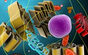
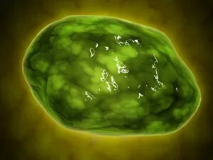
Background imageMembrane Collection: Conceptual image of cell nucleus

Conceptual image of cell nucleus. The cell nucleus acts like the brain of the cell. It helps control eating, movement, and reproduction
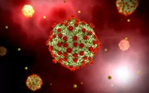
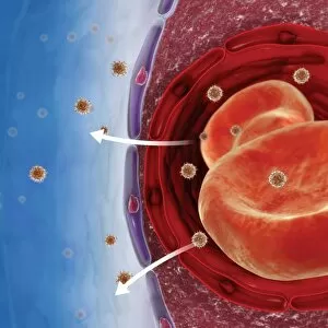
Background imageMembrane Collection: Virus landing on cell membrane

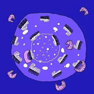
Virus landing on cell membrane

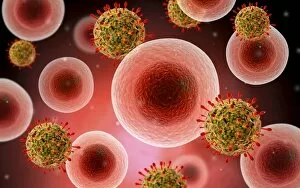
Background imageMembrane Collection: Microscopic view of cell and virus

Microscopic view of cell and virus

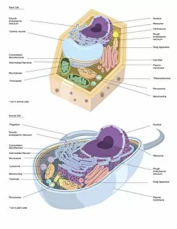
Background imageMembrane Collection: Comparative illustration of plant and animal cell anatomy (with labels)

Comparative illustration of plant and animal cell anatomy (with labels)

Background imageMembrane Collection: Microscopic view of animal cell nucleus

Microscopic view of animal cell nucleus

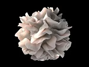
Background imageMembrane Collection: Artistic rendering of the surface of a human dendritic cell

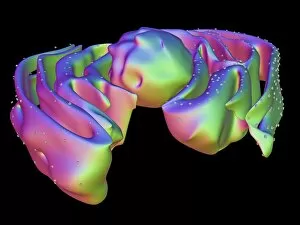
Artistic rendering of the surface of a human dendritic cell illustrating the unexpected discovery of sheet-like processes that fold back onto the membrane surface

Background imageMembrane Collection: Conceptual image of lysosome

Conceptual image of lysosome. Lysosomes are cellular organelles that contain acid hydrolase enzymes that break down waste materials and cellular debris

Background imageMembrane Collection: Microscopic view of HIV virus

Microscopic view of HIV virus

Background imageMembrane Collection: Surface of HIV infected microphage

Surface of HIV infected microphage
3D representation of the surface and interior of an HIV-infected macrophage obtained using newly developed tools for 3D imaging using ion-abrasion scanning electron microscopy

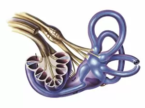
Background imageMembrane Collection: Cochlea detail with vestibulocochlear nerve

Cochlea detail with vestibulocochlear nerve

Background imageMembrane Collection: Red blood cells with white blood cells

Red blood cells with white blood cells

Background imageMembrane Collection: External auditory canal of human ear

External auditory canal of human ear

Background imageMembrane Collection: Picture No. 10900915

Picture No. 10900915
Giant Kingfisher (Megaceryle maximus) Date:

Background imageMembrane Collection: Illustration of leg and hand muscles, synovial membrane or synovium

Illustration of leg and hand muscles, synovial membrane or synovium
Medicine: Human anatomy, leg and hand muscles, synovial membrane, or synovium. Drawing

Background imageMembrane Collection: Illustration of chest, muscles, serous membranes and ribcage

Illustration of chest, muscles, serous membranes and ribcage
Medicine: Human anatomy, chest, muscles, serous membranes and ribcage

Background imageMembrane Collection: Illustration of meninges

Illustration of meninges
Medicine: Human anatomy, meninges. Drawing

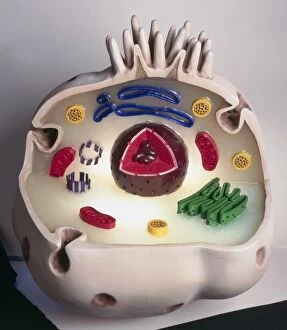
Background imageMembrane Collection: Model of animal cell, including cell nucleus, golgi body, lysosomes, centrioles, mitochondria

Model of animal cell, including cell nucleus, golgi body, lysosomes, centrioles, mitochondria, endoplasmic reticulum, ribosomes, cytoplasm, vesicles, thin plasma membrane

Background imageMembrane Collection: Digital composite showing process of removing waste from human kidney using haemodialysis

Digital composite showing process of removing waste from human kidney using haemodialysis

Background imageMembrane Collection: Mans hands cutting membranes from chicken livers, ground thyme and chopped garlic nearby, close-up

Mans hands cutting membranes from chicken livers, ground thyme and chopped garlic nearby, close-up

Background imageMembrane Collection: Pulling away white membrane from beef tenderloin, using knife, close-up

Pulling away white membrane from beef tenderloin, using knife, close-up

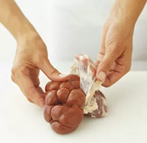
Background imageMembrane Collection: Using fingers to peel membrane off calfs kidney

Using fingers to peel membrane off calfs kidney

Background imageMembrane Collection: Slitting membrane between ribs on neck of lamb, close-up

Slitting membrane between ribs on neck of lamb, close-up

Background imageMembrane Collection: Removing tough membrane from beef fillet

Removing tough membrane from beef fillet

Background imageMembrane Collection: Pulling away white fat (suet) surrounding whole kidney

Pulling away white fat (suet) surrounding whole kidney

Background imageMembrane Collection: Peeling off the outer membrane of lambs kidney, close-up

Peeling off the outer membrane of lambs kidney, close-up

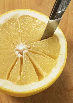
Background imageMembrane Collection: Serrated knife making incisions alongside the membrane lines of half a grapefruit

Serrated knife making incisions alongside the membrane lines of half a grapefruit

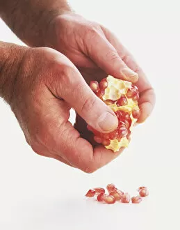
Background imageMembrane Collection: Pomegranate seeds being prised away the membrane, close up

Pomegranate seeds being prised away the membrane, close up

Background imageMembrane Collection: Peeling membrane off whole kidney

Peeling membrane off whole kidney

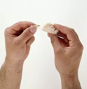
Background imageMembrane Collection: Removing membrane from a scallop, close-up

Removing membrane from a scallop, close-up

Background imageMembrane Collection: Using fingers to pull outer membrane from sweetbread

Using fingers to pull outer membrane from sweetbread

Background imageMembrane Collection: Using a trowel to plant through a horticultural membrane, close-up

Using a trowel to plant through a horticultural membrane, close-up

Background imageMembrane Collection: Cutting horticultural membrane with scissors, close-up

Cutting horticultural membrane with scissors, close-up

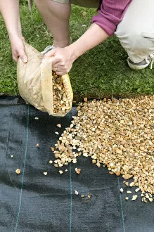
Background imageMembrane Collection: Pouring gravel over permeable membrane to form base of a herb path

Pouring gravel over permeable membrane to form base of a herb path

Background imageMembrane Collection: Frogspawn, small black seeds in jelly-like membrane sack, close-up shot

Frogspawn, small black seeds in jelly-like membrane sack, close-up shot

Background imageMembrane Collection: Chloroplast, artwork

Chloroplast, artwork
Chloroplast, computer artwork

Background imageMembrane Collection: Mitochondria, artwork

Mitochondria, artwork
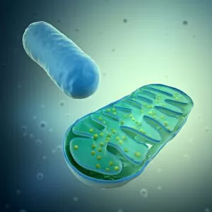
Mitochondria, computer artwork

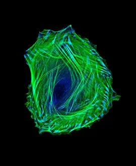
Background imageMembrane Collection: Embryonic smooth muscle cell C018 / 8595

Embryonic smooth muscle cell C018 / 8595
Embryonic smooth muscle cell, immunofluorescence micrograph. Actin filaments are green, the protein vinculin is light blue. Actin is part of the cytoskeleton

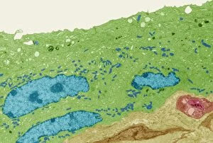
Background imageMembrane Collection: Bladder lining, TEM C014 / 1473

Bladder lining, TEM C014 / 1473
Bladder lining. Transmission electron micrograph (TEM) of a section through the epithelial cells that line of a human bladder, showing large flat vesicles (large, blue)

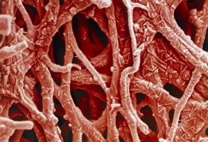
Background imageMembrane Collection: Coloured SEM of internal membrane of hen eggshell

Coloured SEM of internal membrane of hen eggshell
Eggshell membrane. Coloured scanning electron micrograph (SEM) of the internal membrane from the shell of a hens egg. Several membranes are found around the embryo in a birds egg

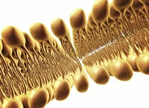
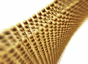
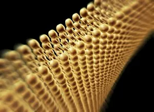
Background imageMembrane Collection: Cell membrane lipid bilayer, artwork F007 / 1479

Cell membrane lipid bilayer, artwork F007 / 1479
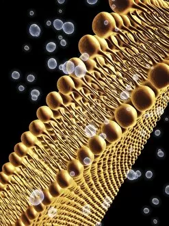
Phospholipid bilayer. Computer artwork of the phospholipid bilayer that forms the membrane around all living cells. The cell membrane is made of phospholipid molecules

Background imageMembrane Collection: Cell membrane lipid bilayer, artwork F007 / 1480

Cell membrane lipid bilayer, artwork F007 / 1480
Phospholipid bilayer. Computer artwork of the phospholipid bilayer that forms the membrane around all living cells. The cell membrane is made of phospholipid molecules

Background imageMembrane Collection: Cell membrane lipid bilayer, artwork F007 / 1478

Cell membrane lipid bilayer, artwork F007 / 1478
Phospholipid bilayer. Computer artwork of the phospholipid bilayer that forms the membrane around all living cells. The cell membrane is made of phospholipid molecules

Background imageMembrane Collection: Cell membrane lipid bilayer, artwork F007 / 1475

Cell membrane lipid bilayer, artwork F007 / 1475
Phospholipid bilayer. Computer artwork of the phospholipid bilayer that forms the membrane around all living cells. The cell membrane is made of phospholipid molecules

Background imageMembrane Collection: Cell membrane lipid bilayer, artwork F007 / 1474

Cell membrane lipid bilayer, artwork F007 / 1474
Phospholipid bilayer. Computer artwork of the phospholipid bilayer that forms the membrane around all living cells. The cell membrane is made of phospholipid molecules

Background imageMembrane Collection: Cell membrane lipid bilayer, artwork F007 / 1473

Cell membrane lipid bilayer, artwork F007 / 1473
Phospholipid bilayer. Computer artwork of the phospholipid bilayer that forms the membrane around all living cells. The cell membrane is made of phospholipid molecules

Background imageMembrane Collection: Wnt signalling pathways, illustration C018 / 0917

Wnt signalling pathways, illustration C018 / 0917
Wnt signalling pathways, illustration. Wnt signalling pathways are three separate pathways that pass signals from outside a cell to inside the cell

Background imageMembrane Collection: Nucleus and endoplasmic reticulum F006 / 9196

Nucleus and endoplasmic reticulum F006 / 9196
Computer artwork showing part of a human or eukaryotic cell. In the middle the nucleus which has a membrane with nuclear pores. Inside the nucleus is the DNA


For sale as Licensed Images

Choose your image, Select your licence and Download the media

The intricate anatomy of the human ear is beautifully depicted in this lithograph, published in 1876. One notable feature highlighted here is the membrane, which plays a crucial role in transmitting sound waves to our auditory system. Moving on to cellular structures, the rough endoplasmic reticulum (ER) takes center stage under a transmission electron microscope (TEM). This network of membranes within cells is responsible for protein synthesis and transport. Artwork showcasing different cell types also emphasizes the significance of membranes. From the delicate cell membrane itself, represented by artwork C013 / 7467, to mitochondria seen through TEM imaging - these organelles possess their own unique membranes that regulate various cellular functions. Intriguingly, even chloroplasts have their own distinct membrane structure as revealed by artwork dedicated to studying photosynthesis. These specialized organelles found in plants are responsible for converting sunlight into energy. Beyond biology, membranes find relevance elsewhere too. Think about damp-proofing measures taken in houses – membranes act as barriers against moisture infiltration and protect our living spaces from potential damage. However, not all mentions of they are positive. Bacterial meningitis can be detected through MRI scans where inflammation affects the protective brain meninges' integrity. Understanding how pathogens breach these defensive layers helps diagnose and treat such infections effectively. Nature's wonders also exhibit fascinating adaptations involving membranes; take Plecotus sp. , commonly known as long-eared bats with their remarkable hearing abilities thanks to specialized ear membrane structures aiding echolocation skills. Zooming into finer details under TEM again reveals eye muscles' intricate arrangement (TEM C014 / 1468), highlighting how well-organized muscle fibers rely on precise membranous connections for coordinated movement and vision control. Lastly, let's not forget intestinal microvilli observed through TEM – finger-like projections covered by plasma membrane lining our intestines play a vital role in nutrient absorption during digestion processes. From ancient lithographs to modern imaging techniques, the significance of membranes spans across various fields.